Jason Langley1, Daniel E Huddleston2, Sumanth Dara3, Ilana Bennett4, and Xiaoping P Hu1,3
1Center for Advanced Neuroimaging, University of California Riverside, Riverside, CA, United States, 2Department of Neurology, Emory University, Atlanta, GA, United States, 3Department of Bioengineering, University of California Riverside, Riverside, CA, United States, 4Department of Psychology, University of California Riverside, Riverside, CA, United States
1Center for Advanced Neuroimaging, University of California Riverside, Riverside, CA, United States, 2Department of Neurology, Emory University, Atlanta, GA, United States, 3Department of Bioengineering, University of California Riverside, Riverside, CA, United States, 4Department of Psychology, University of California Riverside, Riverside, CA, United States
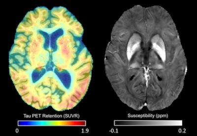
We examine the impact of
APOE-ε4 status on
cortical iron, cortical microstructure, and tau-PET signal in MCI. Our findings suggest that APOE-ε4 allele increases the risk of accumulating tau and iron, which in turn leads to degradation of
cortical tissue microstructure.


