Christopher W Roy1, Leonor Alamo1, Estelle Tenisch1, John Heerfordt1,2, Milan Prsa3, Meritxell Bach Cuadra1,4,5, Davide Piccini1,2, Jérôme Yerly1,4, and Matthias Stuber1,4
1Radiology, Lausanne University Hospital (CHUV) and University of Lausanne (UNIL), Lausanne, Switzerland, 2Advanced Clinical Imaging Technology, Siemens Healthcare, Lausanne, Switzerland, 3Division of Pediatric Cardiology, Department Woman-Mother-Child, Lausanne University Hospital (CHUV) and University of Lausanne (UNIL), Lausanne, Switzerland, 4Center for Biomedical Imaging (CIBM), Lausanne, Switzerland, 5Signal Processing Laboratory 5 (LTS5), Ecole Polytechnique Fédérale de Lausanne (EPFL), Lausanne, Switzerland
1Radiology, Lausanne University Hospital (CHUV) and University of Lausanne (UNIL), Lausanne, Switzerland, 2Advanced Clinical Imaging Technology, Siemens Healthcare, Lausanne, Switzerland, 3Division of Pediatric Cardiology, Department Woman-Mother-Child, Lausanne University Hospital (CHUV) and University of Lausanne (UNIL), Lausanne, Switzerland, 4Center for Biomedical Imaging (CIBM), Lausanne, Switzerland, 5Signal Processing Laboratory 5 (LTS5), Ecole Polytechnique Fédérale de Lausanne (EPFL), Lausanne, Switzerland
A novel framework for 3D MRI of the
fetus with retrospective motion compensation is developed enabling high isotropic
resolution imaging of the entire fetus including the brain and heart.
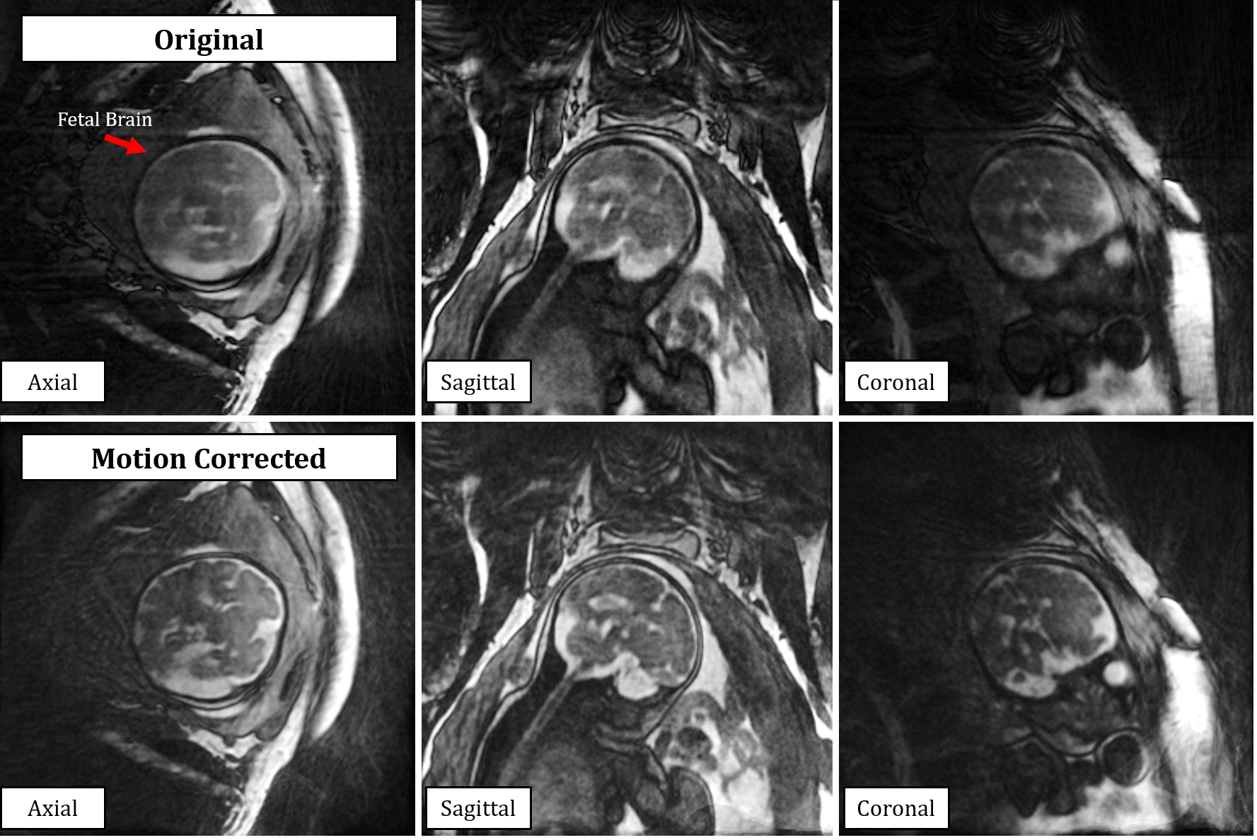
Representative
reconstructions from the proposed motion correction strategy for 3D MRI of the
fetus with isotropic millimetric spatial resolution. The top row shows the originally
acquired (motion-blurred) data in approximate axial, sagittal, and coronal
reformats while the bottom row shows substantial improvement in image quality
and delineation of fetal brain structures after applying retrospective motion correction
to the same data.
Animated
figure depicting motion-resolved reconstructions of free-running 3D fetal MRI
data. A-C) A low spatial resolution real-time image series with 500 ms temporal
resolution demonstrates maternal respiration and gross fetal movement in three
perpendicular planes. D-F) High spatial resolution images created from unique
motion states identified in A) shown in the same views centered on the brain, allowing
for co-registration to “stabilize” anatomical features of interest (G-I).