E.F. Meliadò1,2,3, A.J.E. Raaijmakers1,2,4, M. Maspero2,5, M.H.F. Savenije2,5, P.R. Luijten1, and C.A.T. van den Berg2,5
1Department of Radiology, University Medical Center Utrecht, Utrecht, Netherlands, 2Computational Imaging Group for MR diagnostics & therapy, Center for Image Sciences, University Medical Center Utrecht, Utrecht, Netherlands, 3Tesla Dynamic Coils BV, Zaltbommel, Netherlands, 4Biomedical Image Analysis, Dept. Biomedical Engineering, Eindhoven University of Technology, Eindhoven, Netherlands, 5Department of Radiotherapy, Division of Imaging & Oncology, University Medical Center Utrecht, Utrecht, Netherlands
1Department of Radiology, University Medical Center Utrecht, Utrecht, Netherlands, 2Computational Imaging Group for MR diagnostics & therapy, Center for Image Sciences, University Medical Center Utrecht, Utrecht, Netherlands, 3Tesla Dynamic Coils BV, Zaltbommel, Netherlands, 4Biomedical Image Analysis, Dept. Biomedical Engineering, Eindhoven University of Technology, Eindhoven, Netherlands, 5Department of Radiotherapy, Division of Imaging & Oncology, University Medical Center Utrecht, Utrecht, Netherlands
The deep-learning based Bayesian approach allows
accurate local SAR estimations and returns reliable feedbacks on the confidence/uncertainty
of the estimates.

Figure
4: Generalization Analysis: Ground-Truth local SAR distribution,
predicted
local SAR distributions ($$$\widehat{\mu}$$$), absolute error and estimated
uncertainty ($$$\widehat{\sigma}$$$), for: A
body array with 8 fractionated dipoles
placed on the generic body model Duke10 (without body profile deformation) for
prostate imaging (A) and liver imaging (B); Three head
arrays, one with 8 fractionated dipoles (C), one with 8 oblique fractionated
dipoles11 (D) and one with 8
rectangular loops12
(E).
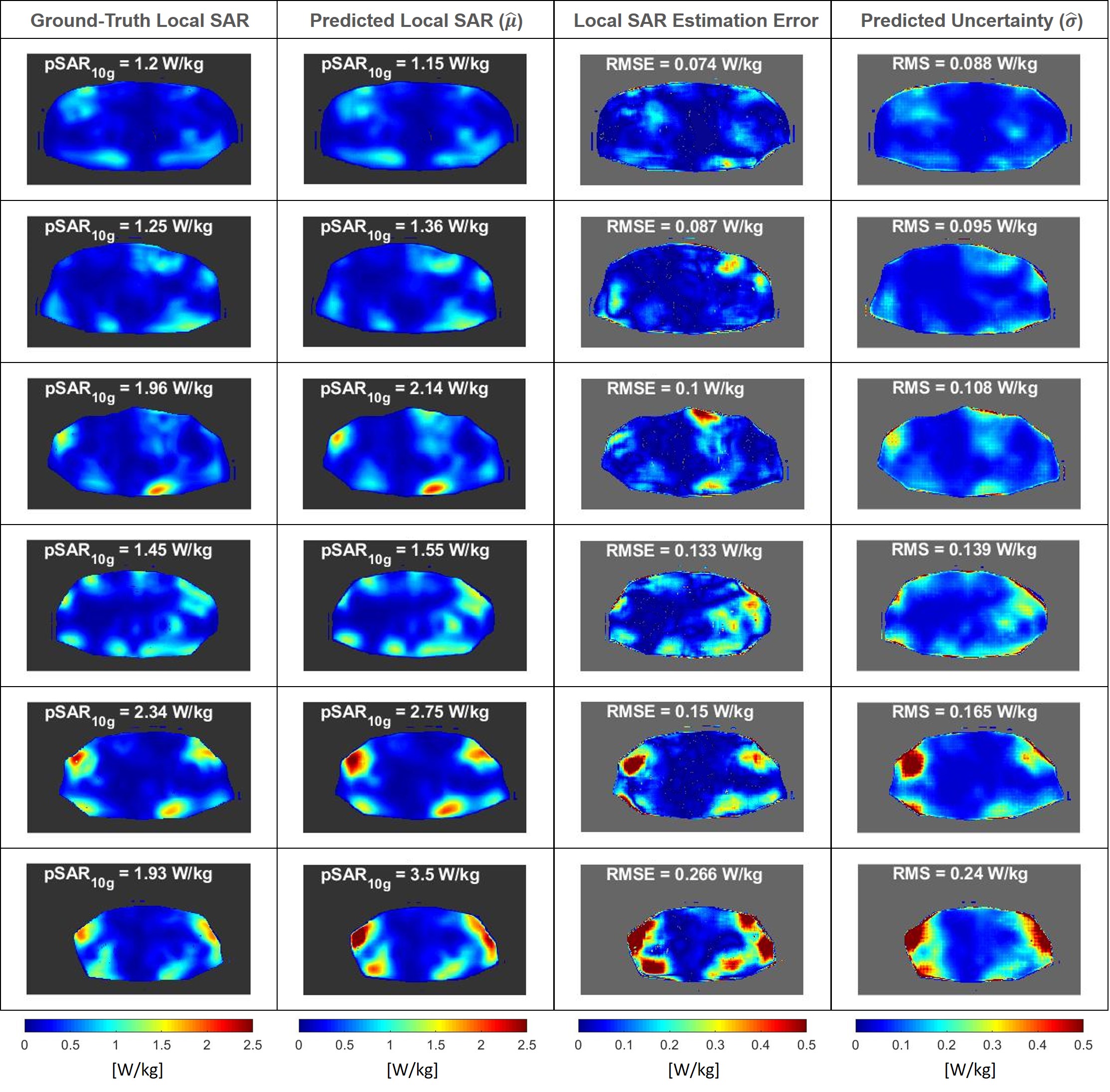
Figure 1: Six
example results of
the 3-Fold Cross-Validation: Ground-Truth
local SAR distribution (first column), predicted local SAR distributions ($$$\widehat{\mu}$$$, second column), absolute error (third column) and estimated uncertainty ($$$\widehat{\sigma}$$$, fourth column). On top are reported the peak
local SAR (pSAR10g), the root-mean-square error
(RMSE) of the absolute error, and the root-mean-square (RMS) of the estimated
uncertainty.