Charlotte E Buchanan1, Hao Li2, Fabio Nery3, Alexander J Daniel1, Joao De Sousa4, Steven Sourbron4, Andrew Priest2,5, David Thomas6,7,8, and Susan T Francis1
1Sir Peter Mansfield Imaging Centre, University of Nottingham, Nottingham, United Kingdom, 2Department of Radiology, University of Cambridge, Cambridge, United Kingdom, 3Great Ormond Street Institute of Child Health, University College London, London, United Kingdom, 4University of Sheffield, Sheffield, United Kingdom, 5Department of Radiology, Addenbrooke's Hospital, Cambridge, United Kingdom, 6Neuroradiological Academic Unit, UCL Queen Square Institute of Neurology, UCL, London, United Kingdom, 7Dementia Research Centre, UCL Queen Square Institute of Neurology, UCL, London, United Kingdom, 8Wellcome Centre for Human Neuroimaging, UCL Queen Square Institute of Neurology, UCL, London, United Kingdom
1Sir Peter Mansfield Imaging Centre, University of Nottingham, Nottingham, United Kingdom, 2Department of Radiology, University of Cambridge, Cambridge, United Kingdom, 3Great Ormond Street Institute of Child Health, University College London, London, United Kingdom, 4University of Sheffield, Sheffield, United Kingdom, 5Department of Radiology, Addenbrooke's Hospital, Cambridge, United Kingdom, 6Neuroradiological Academic Unit, UCL Queen Square Institute of Neurology, UCL, London, United Kingdom, 7Dementia Research Centre, UCL Queen Square Institute of Neurology, UCL, London, United Kingdom, 8Wellcome Centre for Human Neuroimaging, UCL Queen Square Institute of Neurology, UCL, London, United Kingdom
We
have developed a multiparametric renal MRI protocol, harmonised across GE,
Philips and Siemens 3T scanners. This
comprises B0 and B1 mapping, diffusion weighted imaging
(DWI), T1, T2 and T2* mapping, phase contrast
(PC-MRI) and volumetric T1- and T2-weighted scans.

Figure 1.Illustration of the
harmonised multiparametric renal MRI protocol
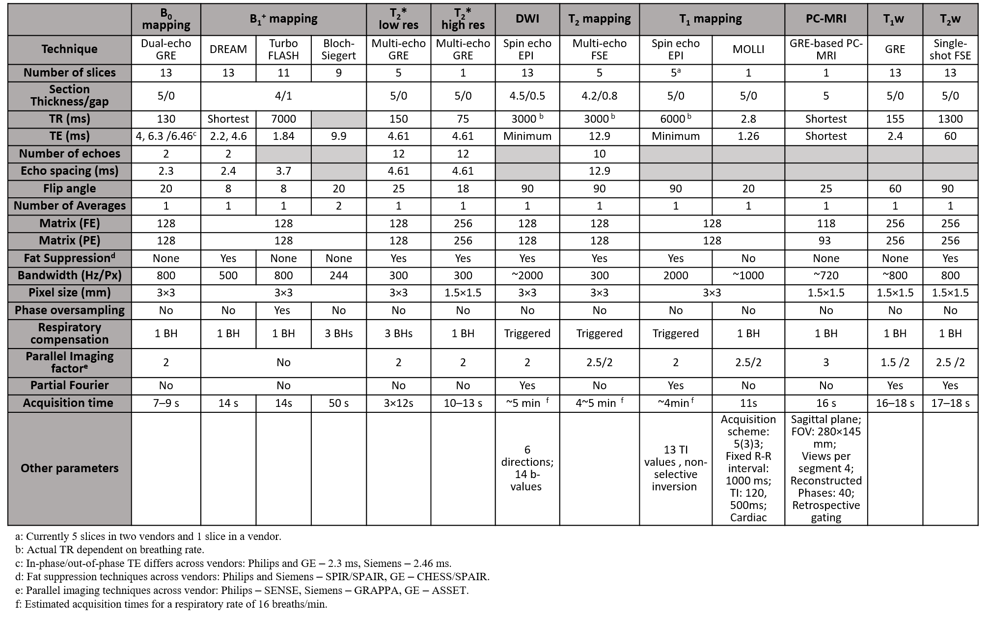
Table
1. The detailed parameters of harmonised multiparametric renal protocols