Neha Atulkumar Singh1, Daniel Gutierrez-Barragan1, Elizabeth de Guzman1, Mauro Uboldi2, Ludovico Coletta1, and Alessandro Gozzi1
1Functional Neuroimaging Laboratory, Istituto Italiano di Tecnologia, Rovereto, Italy, 2Ugo Basile S.r.L., Gemonio, Italy
1Functional Neuroimaging Laboratory, Istituto Italiano di Tecnologia, Rovereto, Italy, 2Ugo Basile S.r.L., Gemonio, Italy
By developing a novel strategy for fMRI connectivity
mapping in awake mice, we identified a possible dynamic signature of
consciousness in this species.
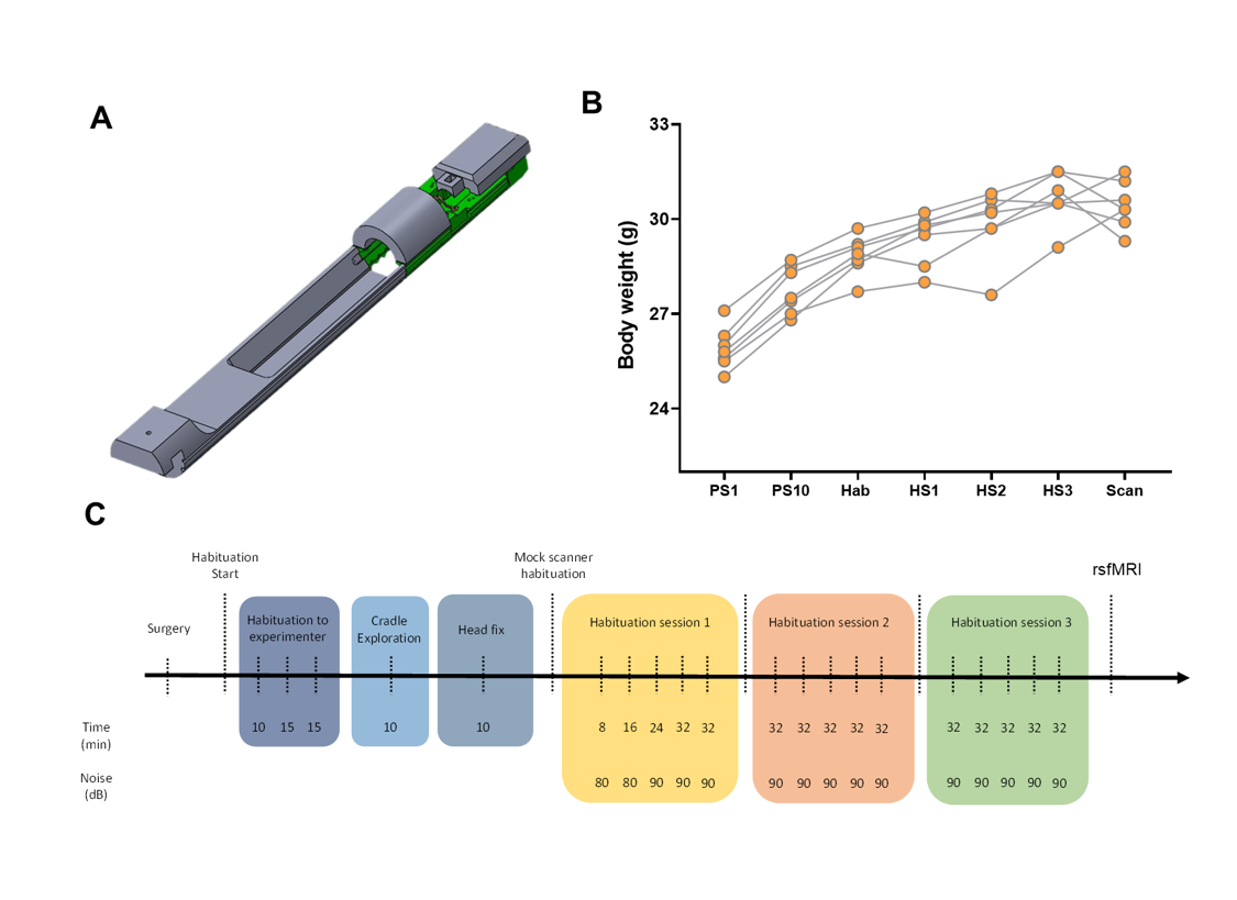
Figure 1. Experimental design for awake rsfMRI acquisitions.
A) Mouse cradle for rsfMRI acquisitions. B) Body weight measurements in 5
representative mice across different sessions. C)
Experimental timeline for habituation protocols [Abv; PS1: post-surgery
day-1, PS10: post-surgery day-10, Hab: Start of habituation, HS1: habituation
session-1, HS2: habituation session-2, HS3: habituation session-3, Scan: rsfMRI
acquisitions]
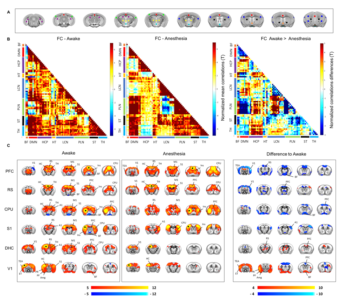
Figure 3. rsfMRI connectivity
networks differ between awake and anesthesia. A) Seed-location for whole-brain connectivity
quantifications in (B). B) Whole-brain group-averaged connectivity matrices for
awake, anaesthesia and mean correlation differences across states. c) rsfMRI
connectivity maps across states, and corresponding difference maps for six
representative networks.