Mada Hashem1,2,3,4,5, Ying Wu1,3,4,5, and Jeff F. Dunn1,2,3,4,5
1Department of Radiology, University of Calgary, Calgary, AB, Canada, 2Biomedical Engineering Graduate Program, University of Calgary, Calgary, AB, Canada, 3Hotchkiss Brain Institute, University of Calgary, Calgary, AB, Canada, 4Experimental Imaging Centre, University of Calgary, Calgary, AB, Canada, 5Cumming School of Medicine, University of Calgary, Calgary, AB, Canada
1Department of Radiology, University of Calgary, Calgary, AB, Canada, 2Biomedical Engineering Graduate Program, University of Calgary, Calgary, AB, Canada, 3Hotchkiss Brain Institute, University of Calgary, Calgary, AB, Canada, 4Experimental Imaging Centre, University of Calgary, Calgary, AB, Canada, 5Cumming School of Medicine, University of Calgary, Calgary, AB, Canada
Cuprizone mice have a loss of white matter and may have
mitochondrial impairment. Multimodality NIRS-MRI revealed an abnormality in
mitochondrial energy production and a reduction in the consumption rate of
oxygen in the cortex.
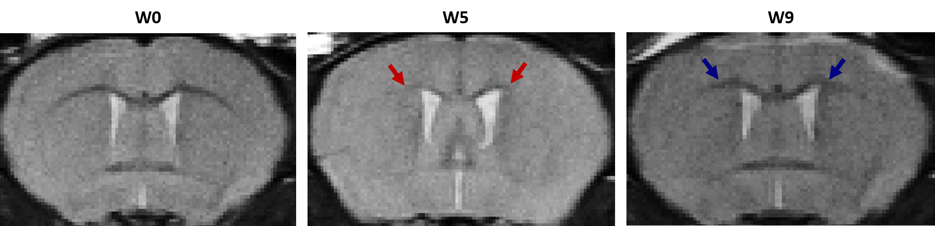
Figure 5: Representative T2-weighted images of a CPZ mouse pre-
(W0), 5 weeks (W5) post- cuprizone exposure, and 4 additional weeks (W9) post
cuprizone exposure termination. A strong gray-white matter contrast which was observed
at baseline decreased at week 5 (red arrows) and increased back (blue arrows)
after recovery (W9). Unlike in the corpus
callosum, this change was not visually obvious in the cerebral cortex.

Figure 1: A reduction in OEF, and CMRO2 in
the cortex of Cuprizone (CPZ) mice but no change in CBF during demyelination. CTRL (black, n=9)
and CPZ (blue, n=11) mice were imaged pre- (W0), 5 weeks (W5) post- cuprizone
exposure, and 4 additional weeks (W9) post cuprizone challenge termination.
Each symbol represents a different mouse (* - p ≤ 0.05, ** - p ≤ 0.01, ***
- p ≤ 0.001).
