Hossam El-Rewaidy1,2, Rui Guo1, and Reza Nezafat1
1Medicine, Beth Israel Deaconess Medical Center and Harvard Medical School, Boston, MA, United States, 2Graduate School of Bioengineering, Department of Computer Science, Technical University of Munich, Munich, Germany
1Medicine, Beth Israel Deaconess Medical Center and Harvard Medical School, Boston, MA, United States, 2Graduate School of Bioengineering, Department of Computer Science, Technical University of Munich, Munich, Germany
A deep artificial neural network (MyoMapNet) enables fast and precise myocardial T1 mapping quantification
from only 4-5 T1-weighted images collected after a
single inversion pulse, leading to shorter scan time and
breath-holds of 4-5 heartbeats.
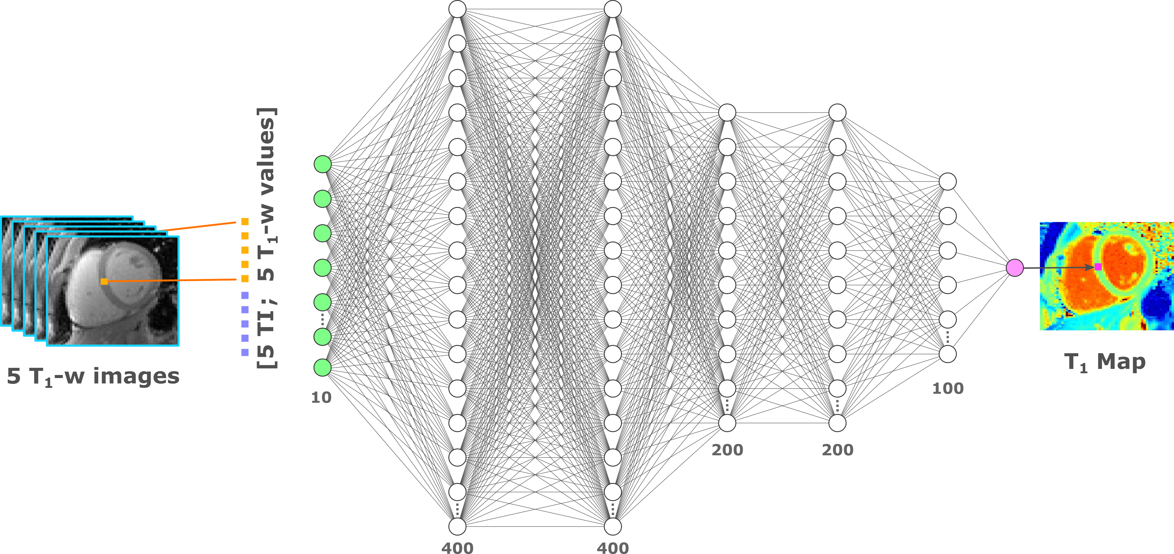
Figure 1. MyoMapNet architecture: MyoMapNet uses a fully-connected neural network for estimating voxel-wise
T1 values from T1-weighted images collected after a single
look-locker inversion pulse. For each voxel, the signal values from 5 T1-weighted images are concatenated
with their corresponding look-locker times and used as the network input (i.e.
10×1) for native T1 mapping. The input values are fed to a
fully-connected network with 5 hidden layers with 400, 400, 200, 200, and 100
nodes each layer, respectively. The output is the estimated T1 value
at each voxel.
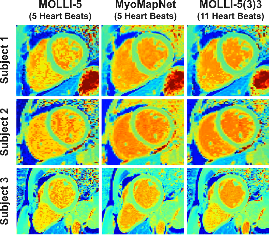
Figure 3. Native T1 maps from three patients, reconstructed using MOLLI-5
(using only 5 T1 weighted images with 3-parameter fitting),
MyoMapNet, and MOLLI-5(3)3 with a 3-parameter fitting model. MoyMapNet yield
maps with more homogenous signal compared to MOLLI-5.