Hanyue Zhou1, Jiayu Xiao2, Debiao Li1,2, Dan Ruan1,3, and Zhaoyang Fan2,4,5
1Bioengineering, University of California, Los Angeles, Los Angeles, CA, United States, 2Cedars-Sinai Medical Center, Los Angeles, CA, United States, 3Radiation Oncology, University of California, Los Angeles, Los Angeles, CA, United States, 4Radiology, University of Southern California, Los Angeles, CA, United States, 5Radiation Oncology, University of Southern California, Los Angeles, CA, United States
1Bioengineering, University of California, Los Angeles, Los Angeles, CA, United States, 2Cedars-Sinai Medical Center, Los Angeles, CA, United States, 3Radiation Oncology, University of California, Los Angeles, Los Angeles, CA, United States, 4Radiology, University of Southern California, Los Angeles, CA, United States, 5Radiation Oncology, University of Southern California, Los Angeles, CA, United States
We developed a deep learning method that utilized 2.5D UNet++, with a loss function contains soft Dice coefficient loss and distance-transform-approximated Hausdorff distance loss. The developed network has further enhanced the segmentation performance across metrics from the baseline.
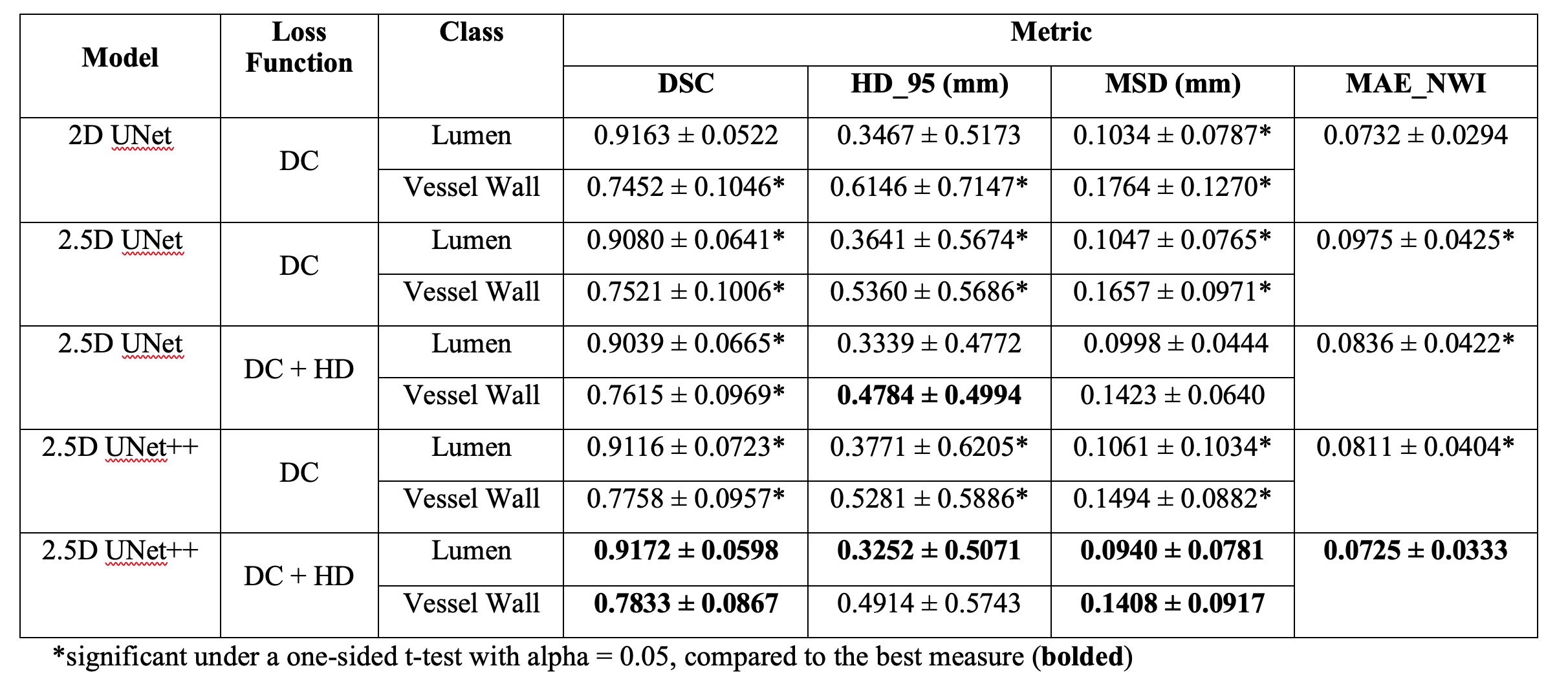
Table I. Quantitative comparison of all models

Fig. 2. Visualization of model performance comparison where the proposed method achieved a better result: dashed block (a) and (b) are two 3-slice examples from a vessel segment. The 1st column is the original consecutive MRI slices (s1, s2, and s3), the 2nd to the last columns show the ground truth and estimated segmentation from each model of the corresponding MRI slice, respectively. Black represents the background, grey represents the vessel wall, and white represents the lumen.