Shinho Cho1, Arani Roy2, Chao Liu2, Djaudat Idiyatullin1, Wei Zhu1, Yi Zhang1, Xiao-Hong Zhu1, Prakash Kara2, Wei Chen1, and Kâmil Uğurbil1
1Center for Magnetic Resonance Research and Department of Radiology, University of Minnesota, Minneapolis, MN, United States, 2Center for Magnetic Resonance Research and Department of Neuroscience, University of Minnesota, Minneapolis, MN, United States
1Center for Magnetic Resonance Research and Department of Radiology, University of Minnesota, Minneapolis, MN, United States, 2Center for Magnetic Resonance Research and Department of Neuroscience, University of Minnesota, Minneapolis, MN, United States
Isotropic high-resolution CBV weighted fMRI, and 2- and 3-photon imaging
studies performed in the cat visual cortex both reveal similar distinctive cortical depth-specific changes in
orientation selectivity and demonstrate that the middle cortical layer is least
selective.
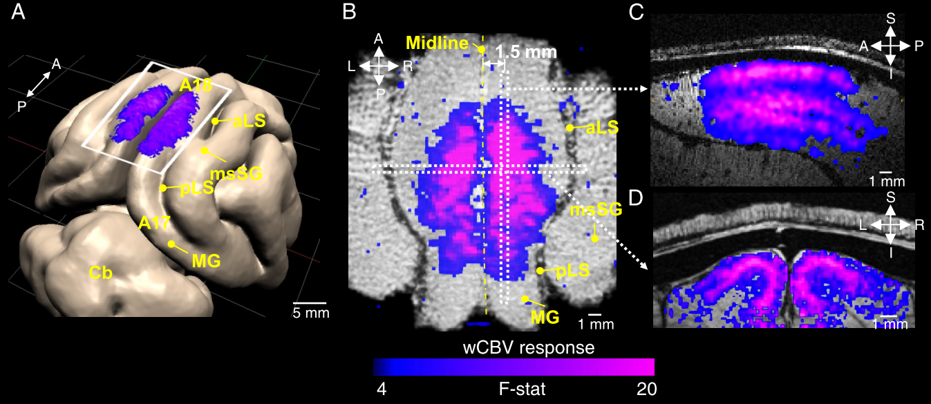
Fig.
1. CBV-weighted (wCBV) activation maps evoked by visual stimuli. (A) Area 18 of cat primary visual cortex; field of view of imaging denoted
by a white rectangular box. F-statistics of wCBV activation induced by 8
orientation gratings presented with blue-purple colors. The pronounced wCBV
activation (P < 0.001) from the
volumetric data displayed on orthogonal anatomical slices: (B) axial, (C) sagittal
and (D)
coronal plane.

Fig. 2. Laminar iso-orientation maps
in cat visual cortex obtained by fMRI. (A) a coronal slice where radial
intracortical veins are seen as dark lines. The yellow lines indicate 7 slices
cut orthogonal to the cortical surface by (post-processed including
interpolation) of the volumetric data. (B) laminar orientation preference
maps seen in these 7 slices. (C) Interpolated depth-dependent
orientation selectivity index (OSI) from wCBV fMRI; individual animals
(gray lines) and group average (black line, n
= 7 cats). Short black vertical lines represent ±1 S.E.M.