Yiming Xiao1,2, Terry M Peters3,4,5, and Ali R Khan3,4,5,6
1PERFORM Centre, Concordia University, Montreal, QC, Canada, 2Computer Science and Software Engineering, Concordia University, Montreal, QC, Canada, 3Robarts Research Institute, Western University, London, ON, Canada, 4Department of Medical Biophysics, Western University, London, ON, Canada, 5School of Biomedical Engineering, Western University, London, ON, Canada, 6The Brain and Mind Institute, Western University, Lonon, ON, Canada
1PERFORM Centre, Concordia University, Montreal, QC, Canada, 2Computer Science and Software Engineering, Concordia University, Montreal, QC, Canada, 3Robarts Research Institute, Western University, London, ON, Canada, 4Department of Medical Biophysics, Western University, London, ON, Canada, 5School of Biomedical Engineering, Western University, London, ON, Canada, 6The Brain and Mind Institute, Western University, Lonon, ON, Canada
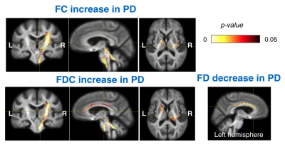
DTI measures and fixel-based analysis revealed strengthened and weakened white matter integrity, which is subject to laterality of motor symptoms in de novo Parkinson’s disease, suggesting both functional degeneration and compensatory network creation.

Figure 1. Investigation of white matter tract changes between Parkinson’s and healthy subjects, using fixel-based analysis (FBA). The white matter tracts that are statistically significant (p<0.05) in fiber cross-section (FC), fiber density (FD) and fiber density and cross-section (FDC) are shown overlaid on a group-averaged template with a heat map. The cross-hair in each sub-figure points at the same location.

Figure 2. Investigation of white matter changes between Parkinson’s and healthy subjects, using voxel-based analysis with fractional anisotropy (FA) and mean diffusivity (MD). The regions that are statistically significant (p<0.05) are shown overlaid on a group-averaged template with a heat map. The cross-hair in each sub-figure points at the same location.