Bhumi Bhusal1, Jason Stockmann2, Azma Mareyam2, John Kirsch2, Lawrence L Wald2, Mark J Nolt1, Joshua Rosenow1, Roberto Lopez-Rosado1, Behzad Elahi1, and Laleh Golestanirad1
1Northwestern University, Chicago, IL, United States, 2Massachusetts General Hospital, Charlestown, MA, United States
1Northwestern University, Chicago, IL, United States, 2Massachusetts General Hospital, Charlestown, MA, United States
7T MRI may be safely performed in patients with deep brain stimulation
implants with careful evaluation of implant and MRI hardware.
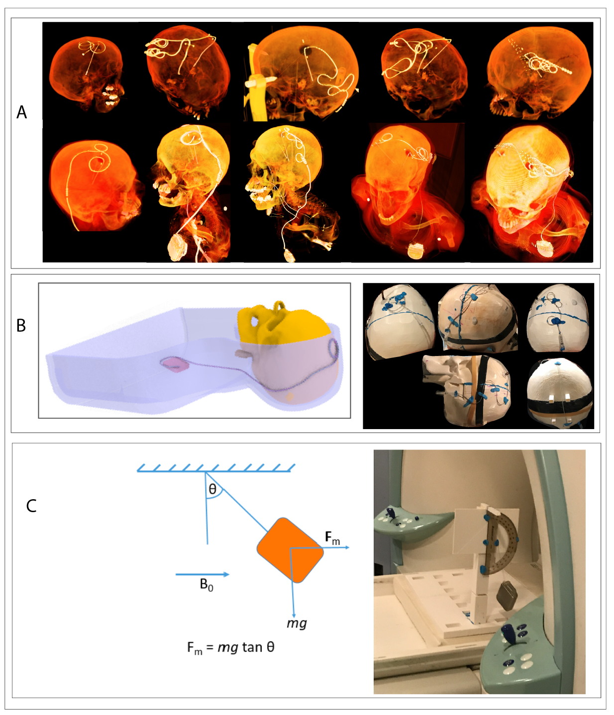
Figure 2. (A) 3D-rendered view of postoperative CT images of DBS
patients showing extracranial trajectories of leads and extensions. (B)
Schematic of the experimental phantom showing a full DBS system as well as
examples of some trajectories used during the RF heating experiments. (C) Setup
for the measurement of the magnetic force on the IPG.
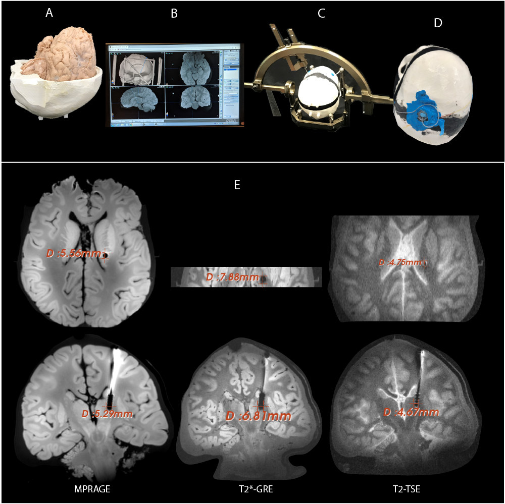
Figure 3. Steps of DBS implantation and artifact measurement. (A)
Formalin fixed cadaveric brain placed inside a 3D printed skull. (B) 1.5T MRI
of the cadaveric brain were transferred to BrainLAB iPlan server for localizing
subthalamic nucleus. (C) Leksell G Frame and stereotactic arc attached to the skull for electrode implantation. (D) Burr hole cover
placed to fixate the lead in position.
(E) Artifact was quantified on transverse and coronal planes for different
imaging sequences.
