Fuyixue Wang1,2, Zijing Dong1,3, Timothy G. Reese1, Lawrence L. Wald1,2, and Kawin Setsompop4,5
1Athinoula A. Martinos Center for Biomedical Imaging, Massachusetts General Hospital, Charlestown, MA, United States, 2Harvard-MIT Health Sciences and Technology, MIT, Cambridge, MA, United States, 3Department of Electrical Engineering and Computer Science, MIT, Cambridge, MA, United States, 4Department of Radiology, Stanford University, Stanford, CA, United States, 5Department of Electrical Engineering, Stanford University, Stanford, CA, United States
1Athinoula A. Martinos Center for Biomedical Imaging, Massachusetts General Hospital, Charlestown, MA, United States, 2Harvard-MIT Health Sciences and Technology, MIT, Cambridge, MA, United States, 3Department of Electrical Engineering and Computer Science, MIT, Cambridge, MA, United States, 4Department of Radiology, Stanford University, Stanford, CA, United States, 5Department of Electrical Engineering, Stanford University, Stanford, CA, United States
This work develops optimized 3D-EPTI whole brain protocols at 1-mm and 0.7-mm isotropic resolutions for rapid quantitative mapping, and demonstrates a high level of repeatability achieved in the derived quantitative maps across brain regions and cortical depths via scan-rescan validation.
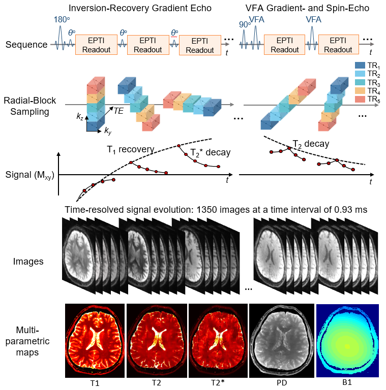
Figure 1. The pulse sequence diagram of 3D EPTI. The signals of inversion recovery gradient echo (IR-GE) and variable flip angle gradient-echo-and-spin-echo (GRASE) are acquired using a spatiotemporal encoding in k-t domain, which encodes a block of the 3D k-t space that later forms a radial-blade after combining the data across different TRs. The radial-blades are with different angulation to create incoherence along time to help with time-resolving ~1350 images at TE increments of ~1ms. The time-resolved images are used to fit quantitative parameters of T1, T2, T2*, PD and B1+ maps.
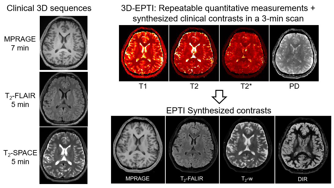
Figure 5. Synthesized multi-contrast images using the 3-minute 1-mm protocol compared with contrast-weighted images acquired by clinical 3D sequences.