Eze Ahanonu1, Zhiyang Fu1,2, Kevin Johnson2, Maria Altbach2,3, and Ali Bilgin1,2,3
1Electrical and Computer Engineering, University of Arizona, Tucson, AZ, United States, 2Medical Imaging, University of Arizona, Tucson, AZ, United States, 3Biomedical Engineering, University of Arizona, Tucson, AZ, United States
1Electrical and Computer Engineering, University of Arizona, Tucson, AZ, United States, 2Medical Imaging, University of Arizona, Tucson, AZ, United States, 3Biomedical Engineering, University of Arizona, Tucson, AZ, United States
An accelerated T1 mapping framework which utilizes
deep learning to estimate T1 using a fraction of the T1 recovery curve (T1RC)
is presented. In vivo experiments demonstrate that the proposed framework can
enable full abdominal coverage within a single BHP.
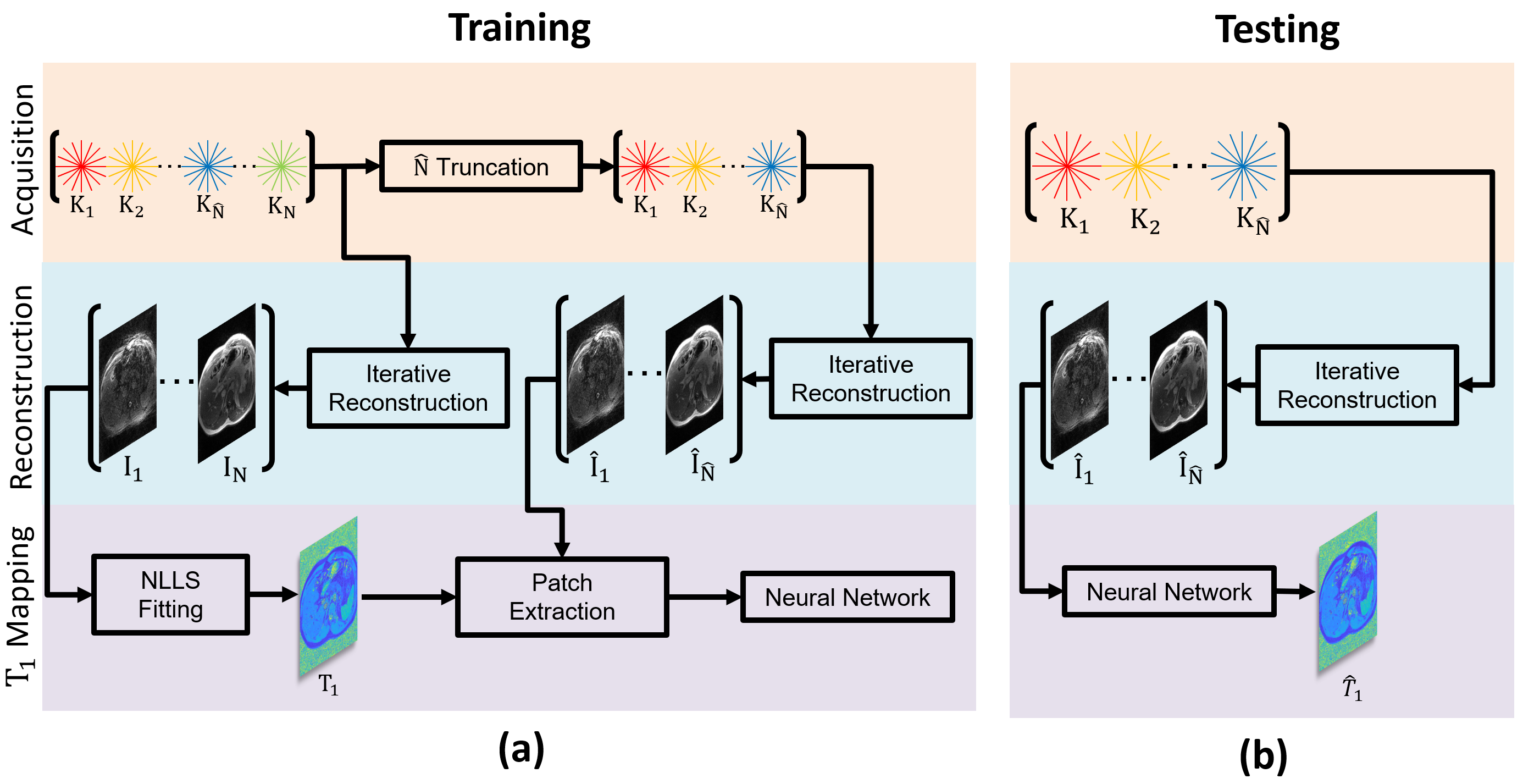
Figure 2: Demonstrating
the training (a) and testing (b) pipelines for deep learning based T1 mapping. In the figure, the pipelines are shown for a
single slice. During training, TI images are obtained with and without
truncation of the acquired k-space data along the T1 recovery curve (T1RC). The
images from the truncated T1RC k-space are used as input and the T1 map from
the full T1RC is used as target.

Figure 5: T1
estimation performance for a subject with a liver lesion for both the proposed DL
framework and NLLS fitting with $$$\hat{N}=8,12,16,20,24$$$ and $$$28$$$ and TIs as
input. In (a) results for three 9x9 pixel ROIs are given. The height of each
bar represents the average T1 value within a given ROI, with error bars
representing the standard deviation. In (b), visual examples are provided for
qualitative evaluation of the two techniques over the liver lesion with varying
TIs.