Carlos Velasco1, Giorgia Milotta1, Alina Hua1, Karl Kunze1,2, Radhouene Neji1,2, Tevfik Ismail1, Claudia Prieto1, and René M. Botnar1
1School of Biomedical Engineering and Imaging Sciences, King's College London, London, United Kingdom, 2MR Research Collaborations, Siemens Healthcare Limited, Frimley, United Kingdom
1School of Biomedical Engineering and Imaging Sciences, King's College London, London, United Kingdom, 2MR Research Collaborations, Siemens Healthcare Limited, Frimley, United Kingdom
The proposed free-breathing accelerated and motion corrected 3D joint T1/T2 sequence, that allows the acquisition of isotropic T1 and T2 maps and complementary water and fat images in ~9 min has been evaluated in patients with cardiovascular disease.
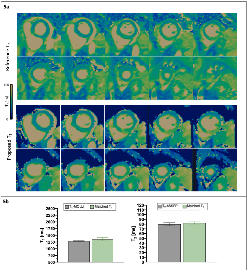
Fig. 5 a) Volumetric SA quantification on the reference (top) and proposed (bottom) T2 maps of Subject #2 obtained from the proposed approach. b) Quantification, for the same subject, inside an ROI manually drawn within the region encompassing the abnormal T1/T2 values (marked with a white arrow in Figure 4). Good correlation for both T1 and T2 between standard and proposed quantification methods is shown.
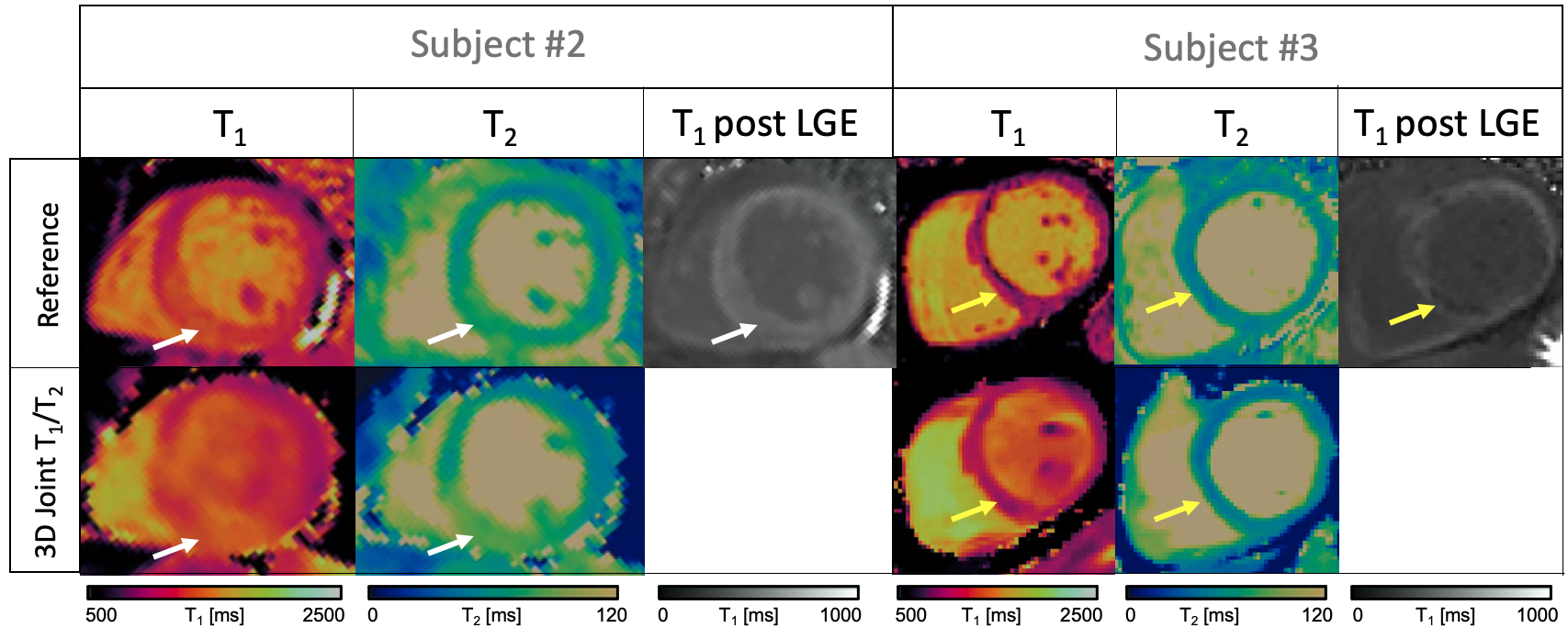
Fig 4. Standard T1 MOLLI and 2D bSSFP (top row) and proposed joint T1/T2 (bottom row) maps obtained two patients with clinical findings. Subject #2, diagnosed with acute myocarditis, presented elevated T1 and T2 values (white arrows). Subject #3 presented with a myocardial infarction in the mid septal region (yellow arrows).