Ingo Hermann1,2, Daiki Tamada3, Scott Reeder3, Lothar Schad2, and Sebastian Weingärtner1
1Magnetic Resonance Systems Lab, Department of Imaging Physics, Delft University of Technology, Delft, Netherlands, 2Computer Assisted Clinical Medicine, Medical Faculty Mannheim, University Heidelberg, Mannheim, Germany, 3Department of Radiology, University of Wisconsin, Madison, WI, United States
1Magnetic Resonance Systems Lab, Department of Imaging Physics, Delft University of Technology, Delft, Netherlands, 2Computer Assisted Clinical Medicine, Medical Faculty Mannheim, University Heidelberg, Mannheim, Germany, 3Department of Radiology, University of Wisconsin, Madison, WI, United States
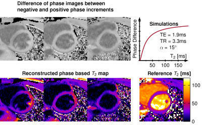
Phase based T2 mapping enables whole heart imaging within one breath hold.

The in vivo signal phase difference is shown for three slices. Simulations were performed for the sequence parameters to calculate from the T2 time estimates from the phase-difference. The reference T2 map and the reconstructed phase based T2 map for three slices are depicted, showing a homogeneous myocardium for two of the three slices. One slice suffered from signal loss in the septal region likely due to B1+ inhomogeneities.

A) Gradient echo acquisition with quadratically increasing phase increments and readout rewinder. B) Simulated phase for a GRE acquisition for different phase increments Φ C) The signal phase is depicted over time for varying T1 and T2 times. D) The signal phase and E) the phase difference for a phase increment of ±2° are shown as a function of T1 and T2. F) The sequence diagram is shown for multiple slices triggered to the end-diastolic phase for positive and negative phase increment. First all slices with positive phase increment are acquired followed by negative phase increment.