Anirban Sengupta1, Arabinda Mishra1, Feng Wang1, Li Min Chen1, and John C. Gore1
1Vanderbilt University Medical Center, Nashville, TN, United States
1Vanderbilt University Medical Center, Nashville, TN, United States
Robust BOLD fMRI fluctuations were detected at
the bilateral intermediate region and gray-commissure region of spinal cord. Selective
disruption of dorsal column white matter tract damages the inter-segmental
connectivity more than the intra-segmental connectivity.

Figure
4. (A) Connectivity matrix at different time-points arranged such that the connectivities from same
community are next to each other. Red boxes on the matrix highlight the communities formed using graph theory
principles. (B)
The
resulting communities are shown in different colors overlaid
on a grid matrix. The
columns of grid matrix represent the seven ROI and the rows represent the five slices/segments. No IR component was observed from
slice 2 and kept white in color. Note community structures change and hence
their number and colors don’t match across time points.
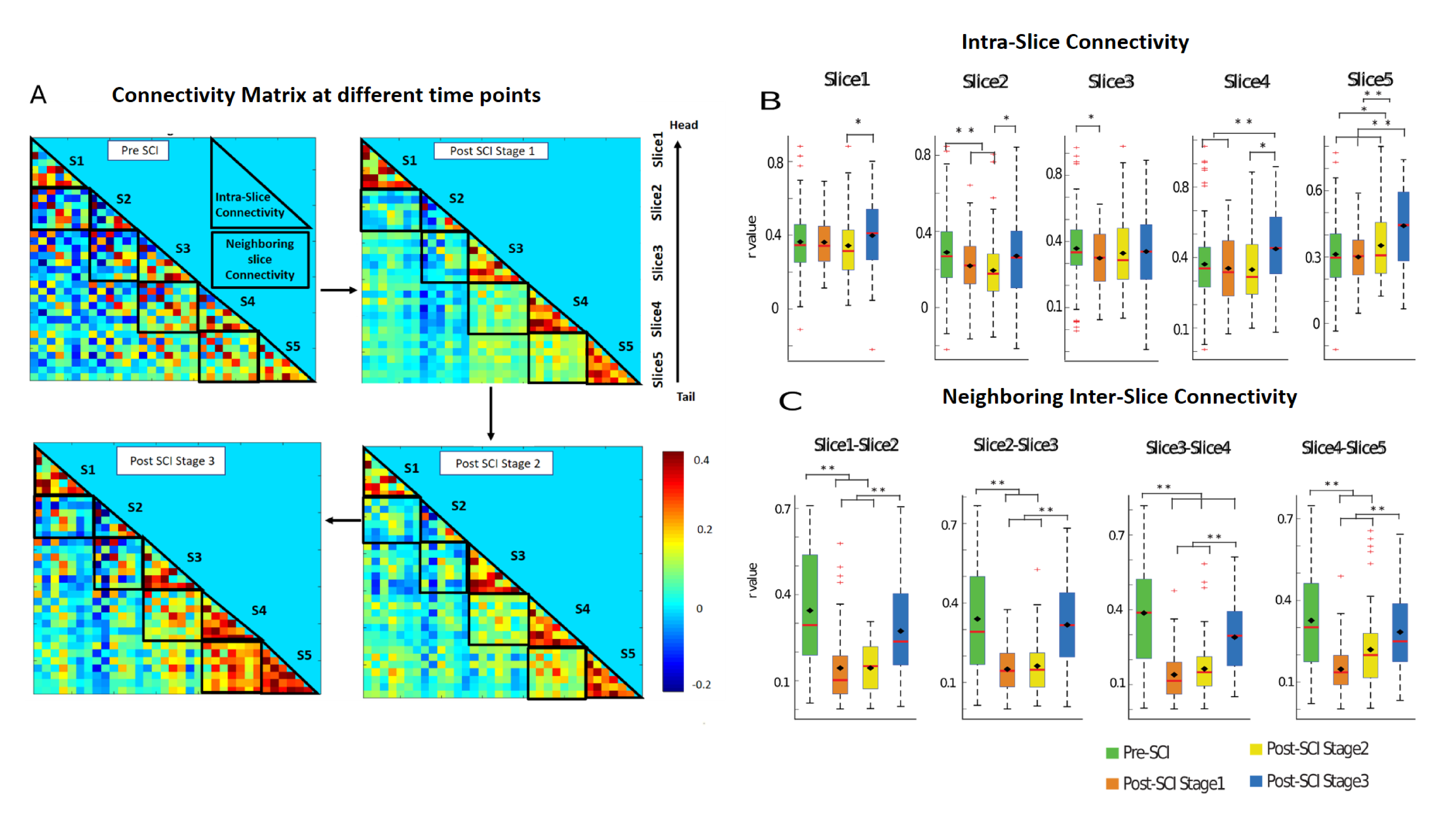
Figure
3: (A) Connectivity
matrix
averaged
over 14 runs of 5 monkeys at
different
time points with intra-slice
and
neighboring inter-slice
connectivities highlighted in black
triangles and black box respectively. Box-plot of (B) intra-slice connectivities and (C)
neighboring
inter-slice connectivities at different time points. Box-plots for
intra-slice and neighboring inter-slice are computed from their corresponding
significant connectivity measures. Significantly different box-plots (Wilcoxon
non-parametric test) are denoted by * (p<0.05) and * * (p<0.01) .
.