Caroline Guglielmetti1,2, Huihui Li3, Lydia M. Le Page1,2, Lauren Y. Shields3, Jeffrey C. Rathmell4, Ken Nakamura3, and Myriam M. Chaumeil1,2
1Department of Physical Therapy and Rehabilitation Science, University of California San Francisco, San Francisco, CA, United States, 2Department of Radiology and Biomedical Sciences, University of California San Francisco, San Francisco, CA, United States, 3Gladstone Institute of Neurological Disease, San Francisco, CA, United States, 4Vanderbilt Center for Immunobiology, Department of Pathology, Microbiology, and Immunology, Vanderbilt University Medical Center, Nashville, TN, United States
1Department of Physical Therapy and Rehabilitation Science, University of California San Francisco, San Francisco, CA, United States, 2Department of Radiology and Biomedical Sciences, University of California San Francisco, San Francisco, CA, United States, 3Gladstone Institute of Neurological Disease, San Francisco, CA, United States, 4Vanderbilt Center for Immunobiology, Department of Pathology, Microbiology, and Immunology, Vanderbilt University Medical Center, Nashville, TN, United States
Deletion of glucose transporter 3 (GLUT3) in a subpopulation of
hippocampal neurons resulted in memory decline in mice. HP 13C
lactate/pyruvate ratio and brain volume were decreased in female mice with a GLUT3
deletion, but not in male mice, while 18F-FDG PET imaging did not
detect changes.

(A)
Representative 13C
spectra showing HP [1-13C]pyruvate and HP [1-13C]lactate
production from a region containing the CA1 area of the hippocampus (red square).
(B) HP [1-13C]lactate/pyruvate ratios were significantly lower
in female GLUT3 cKO compared to GLUT3 WT (p=0.0282). (C) HP [1-13C]lactate/pyruvate
ratios were not significantly different between male GLUT3WT and GLUT3cKO.
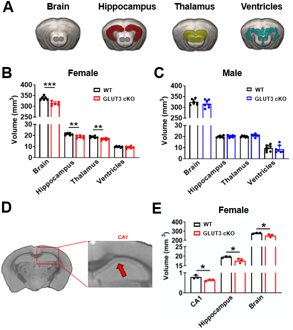
(A) 3D rendering of in vivo T2w MRI showing
the entire brain (grey), hippocampus (red), thalamus (yellow) and ventricle
(blue). (B) In vivo T2w images revealed significantly smaller
brain, hippocampus and thalamus in female GLUT3 cKO compared to WT. (C) No changes were observed in males. (D) Ex vivo T2w images
showing the hippocampus and CA1 region (arrow). CA1, hippocampus and total
brain volumes calculated from ex vivo T2w images were
significantly smaller in female GLUT3 cKO compared to WT.