Dongyue Si1, Shuo Chen1, Daniel A. Herzka2, and Haiyan Ding1
1Center for Biomedical Imaging Research, Department of Biomedical Engineering, Tsinghua University, Beijing, China, 2National Heart, Lung, and Blood Institute, National Institutes of Health, Bethesda, MD, United States
1Center for Biomedical Imaging Research, Department of Biomedical Engineering, Tsinghua University, Beijing, China, 2National Heart, Lung, and Blood Institute, National Institutes of Health, Bethesda, MD, United States
3D T2 mapping techniques enables quantitative detection of
edematous tissue in whole heart. In this study, an accelerated 3D T2
mapping sequence was developed based
on low-rank plus sparsity reconstruction.
Homogeneous whole left ventricular T2 map can
be acquired in single breath-hold.
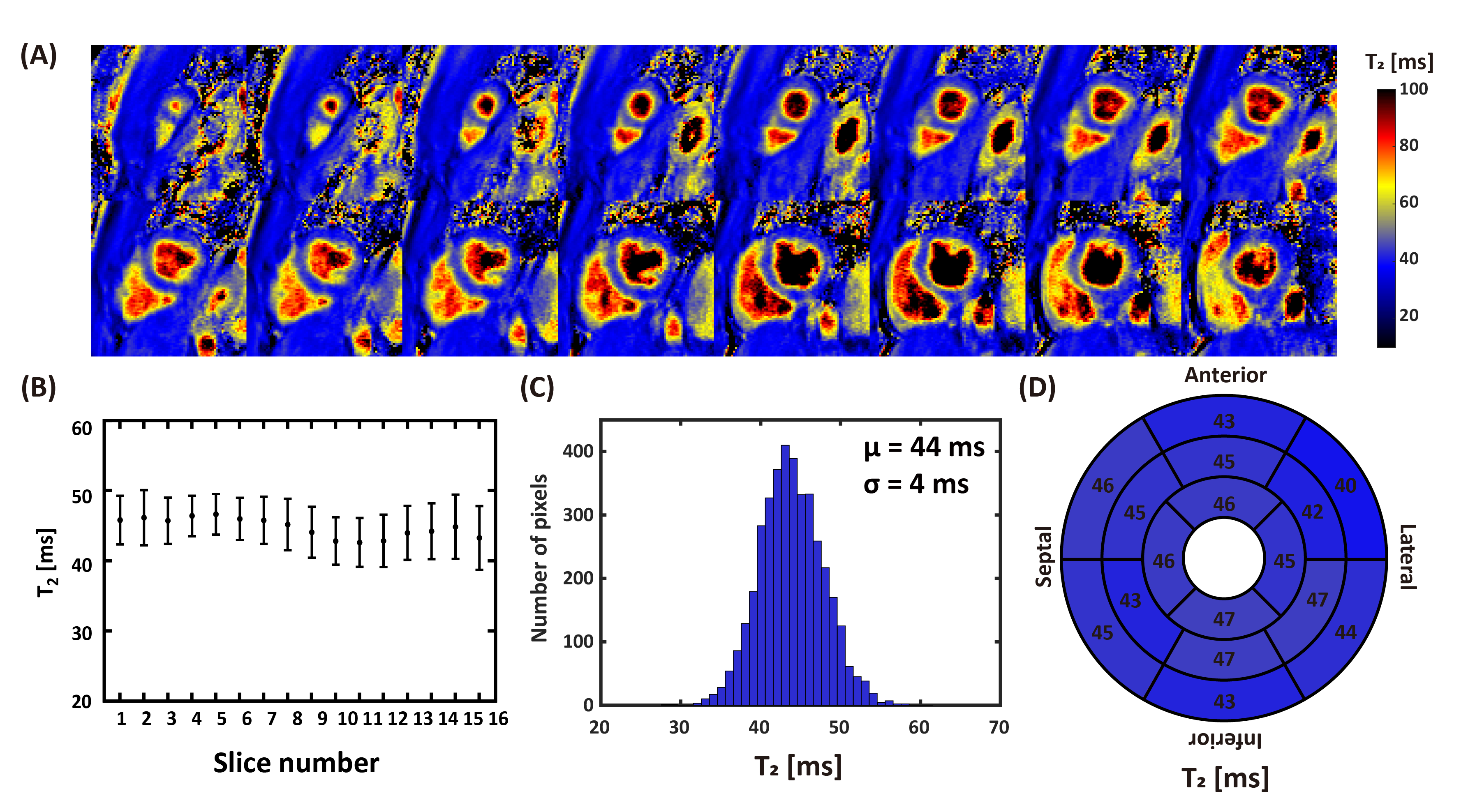
Figure 4. Representative
3D T2 map from one healthy human subject by BH3DT2 sequence. A: 3D whole left ventricle T2 maps from
apex to base. B: mean and standard
deviation of T2 values within every slice. C: Histogram of myocardium T2 over the whole left
ventricle. The mean (μ) and standard deviation (σ) across the whole left
ventricle are shown on the graph. D:
AHA 16-segment bull's-eye plot for showing T2 of each region.
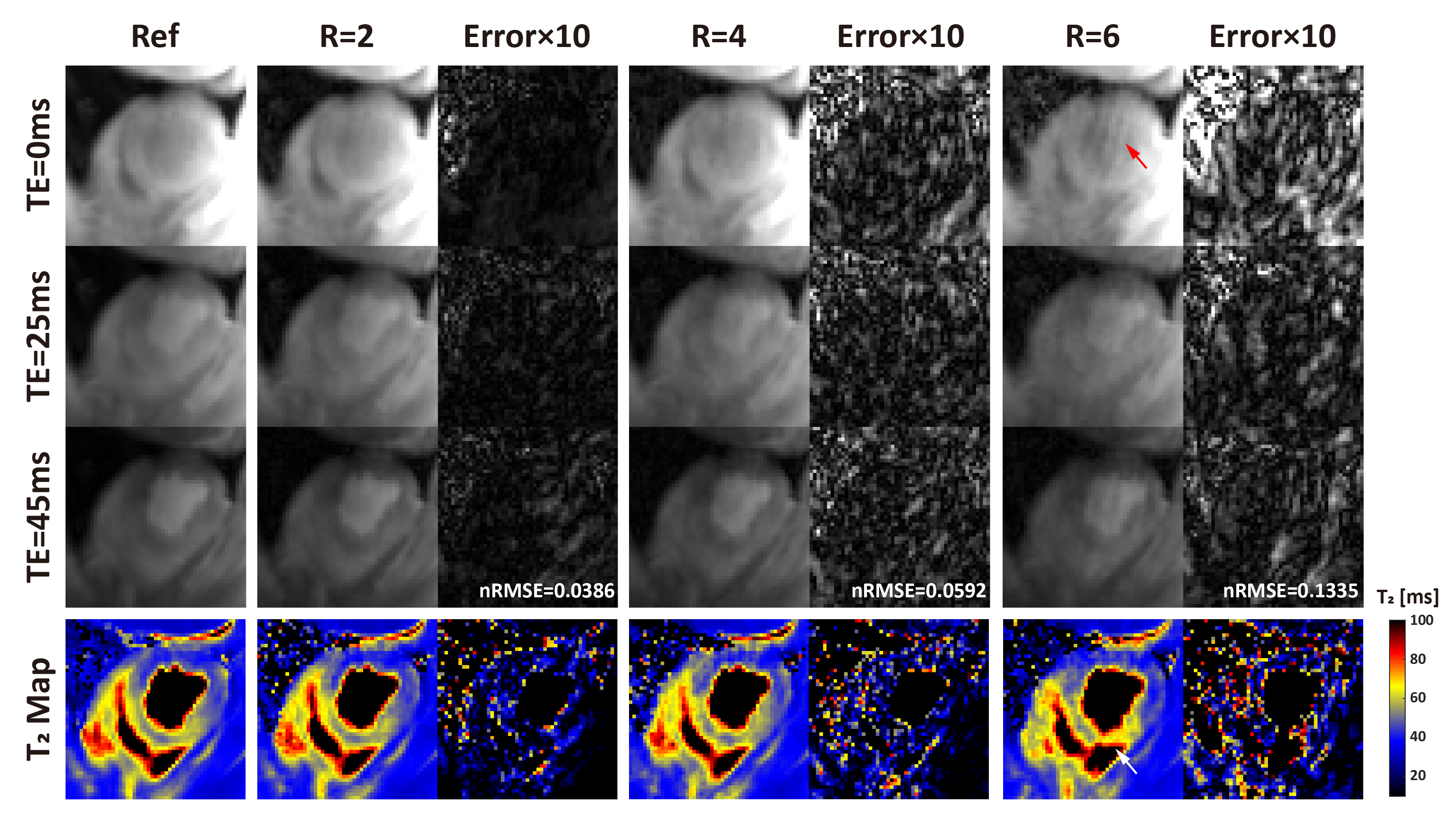
Figure 2. Representative
slice from a swine with acute MI, nRMSE was increased at higher acceleration
factor (R). Image artefacts were subtle at R=2 and 4. At R=6, obvious artefacts
were observed at the blood pool in the T2 weighted image (red arrow),
and septal T2 value was overestimated (white arrow).