Jessica M Winfield1,2, Jennifer C Wakefield1,2, James D Brenton3,4,5, Khalid AbdulJabbar6,7, Antonella Savio8, Susan Freeman9, Erika Pace1,2, Kerryn Lutchman-Singh10, Katherine M Vroobel8, Yinyin Yuan6,7, Susana Banerjee11, Nuria Porta12, Shan E Ahmed Raza6,7,13, and Nandita M deSouza1,2
1MRI Unit, Royal Marsden NHS Foundation Trust, London, United Kingdom, 2Division of Radiotherapy and Imaging, Institute of Cancer Research, London, United Kingdom, 3Cancer Research UK Cambridge Institute, Cambridge, United Kingdom, 4Addenbrooke’s Hospital, Cambridge University Hospitals NHS Foundation Trust, Cambridge, United Kingdom, 5Department of Oncology, University of Cambridge, Cambridge, United Kingdom, 6Centre for Evolution and Cancer, The Institute of Cancer Research, London, United Kingdom, 7Division of Molecular Pathology, The Institute of Cancer Research, London, United Kingdom, 8Department of Pathology, Royal Marsden NHS Foundation Trust, London, United Kingdom, 9Department of Radiology, Addenbrooke’s Hospital, Cambridge University Hospitals NHS Foundation Trust, Cambridge, United Kingdom, 10Swansea Gynaecological Oncology Centre, Swansea Bay University Health Board, Singleton Hospital, Swansea, United Kingdom, 11Gynaecology Unit, Royal Marsden NHS Foundation Trust, Sutton, United Kingdom, 12Clinical Trials and Statistics Unit, The Institute of Cancer Research, London, United Kingdom, 13Department of Computer Science, University of Warwick, Warwick, United Kingdom
1MRI Unit, Royal Marsden NHS Foundation Trust, London, United Kingdom, 2Division of Radiotherapy and Imaging, Institute of Cancer Research, London, United Kingdom, 3Cancer Research UK Cambridge Institute, Cambridge, United Kingdom, 4Addenbrooke’s Hospital, Cambridge University Hospitals NHS Foundation Trust, Cambridge, United Kingdom, 5Department of Oncology, University of Cambridge, Cambridge, United Kingdom, 6Centre for Evolution and Cancer, The Institute of Cancer Research, London, United Kingdom, 7Division of Molecular Pathology, The Institute of Cancer Research, London, United Kingdom, 8Department of Pathology, Royal Marsden NHS Foundation Trust, London, United Kingdom, 9Department of Radiology, Addenbrooke’s Hospital, Cambridge University Hospitals NHS Foundation Trust, Cambridge, United Kingdom, 10Swansea Gynaecological Oncology Centre, Swansea Bay University Health Board, Singleton Hospital, Swansea, United Kingdom, 11Gynaecology Unit, Royal Marsden NHS Foundation Trust, Sutton, United Kingdom, 12Clinical Trials and Statistics Unit, The Institute of Cancer Research, London, United Kingdom, 13Department of Computer Science, University of Warwick, Warwick, United Kingdom
Repeatability of ADC estimates in primary and metastatic tumor sites in epithelial ovarian cancer, and correlation with histopathological metrics (residual tumor and necrosis) after neoadjuvant chemotherapy.
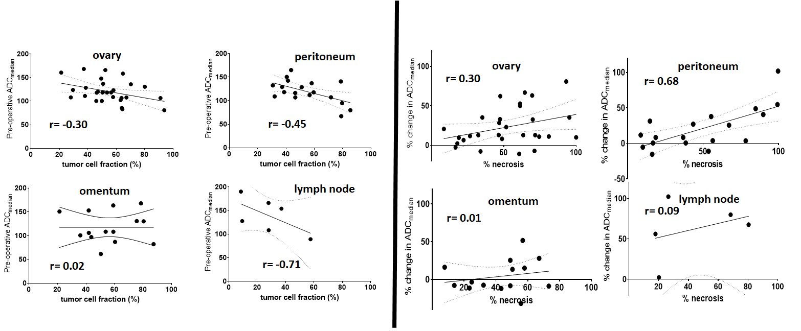
Figure 5: Scatter plots of post-NAC (pre-operative) ADCmedian
vs. tumor cell fraction (left panel) and change in ADCmedian after three or four
cycles of neoadjuvant chemotherapy and percentage necrosis.
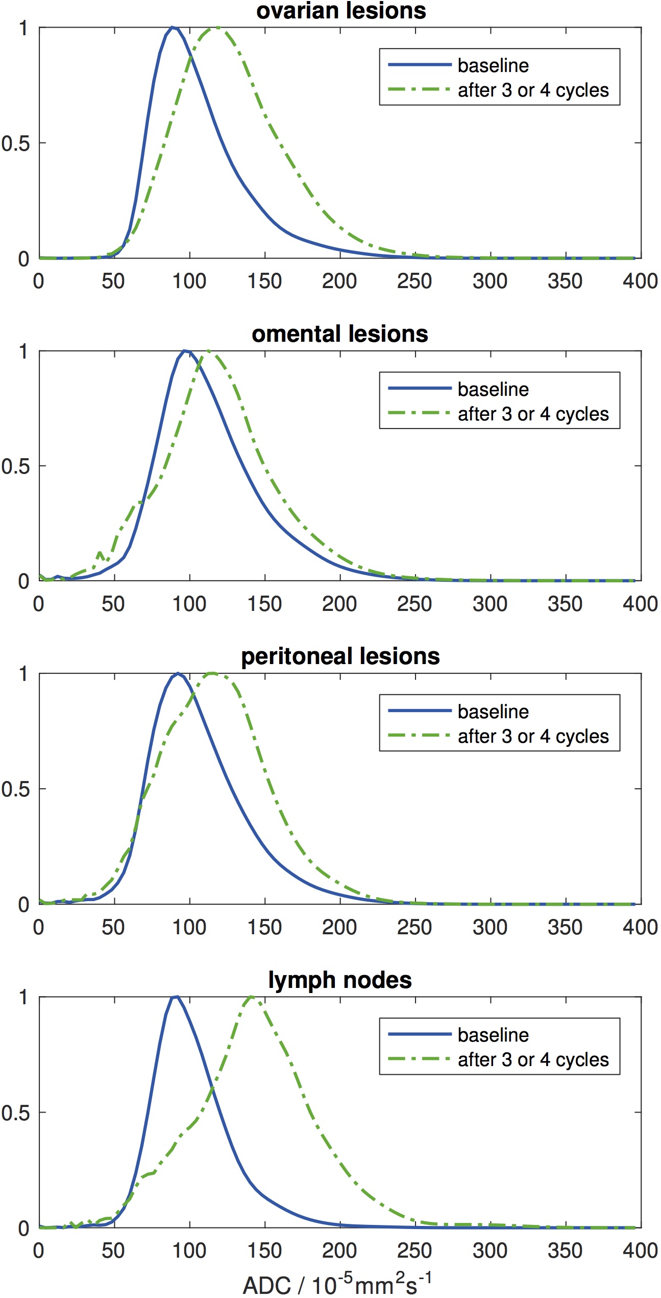
Figure 4: Normalised probability
density functions for ADC estimates from ovarian, omental, and peritoneal
lesions, and lymph nodes at baseline (pre-NAC) and after three or four cycles
of neoadjuvant chemotherapy (post-NAC, pre-operative).