Celine Taglang1, Georgios Batsios1, Mers Tran1, Anne Marie Gillespie1, Hema Artee Luchman2, Russell O Pieper3, Sabrina M Ronen1, and Pavithra Viswanath1
1Radiology and Biomedical Imaging, University of California San Francisco, San Francisco, CA, United States, 2Cell Biology and Anatomy, University of Calgary and Hotchkiss Brain Institute, Calgary, AB, Canada, 3Neurological Surgery, University of California San Francisco, San Francisco, CA, United States
1Radiology and Biomedical Imaging, University of California San Francisco, San Francisco, CA, United States, 2Cell Biology and Anatomy, University of Calgary and Hotchkiss Brain Institute, Calgary, AB, Canada, 3Neurological Surgery, University of California San Francisco, San Francisco, CA, United States
Here, we show that [6,6’-2H]-glucose
non-invasively monitors tumor burden and response to therapy in preclinical low-grade
glioma models at early timepoints prior to alterations in tumor volume, pointing
to its potential to assess pseudoprogression, which is a challenge in glioma
imaging.
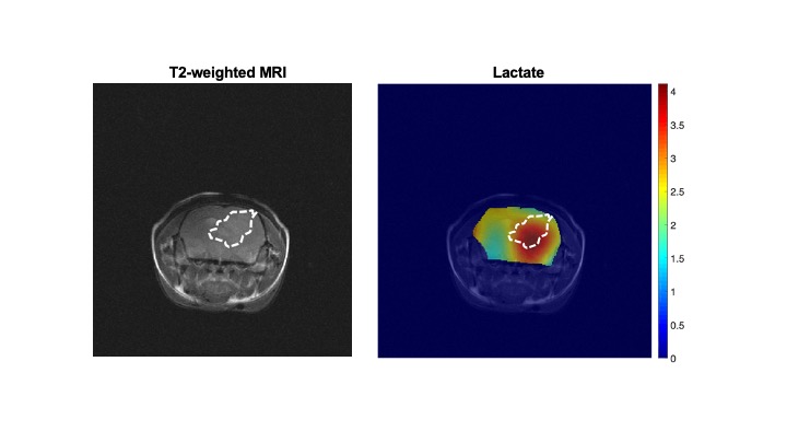
Figure 3. [6,6’-2H]-glucose
flux to lactate is localized to the tumor region in low-grade gliomas in
vivo. Representative metabolic heatmaps from 2D CSI studies in mice
bearing orthotopic BT257 tumor xenografts following injection of a bolus of [6,6’-2H]-glucose.
Left panel shows a representative axial T2-weighted MR image while the right
panel shows a metabolic heatmap of the SNR of lactate produced from [6,6’-2H]-glucose.
