Stephan Kaczmarz1,2, Lena Schmitzer1, Jens Göttler1,2, Kilian Weiss3, Christian Sorg1, Claus Zimmer1, Fahmeed Hyder2, Christine Preibisch1, and Alexander Seiler4
1School of Medicine, Department of Neuroradiology, Technical University of Munich (TUM), Munich, Germany, 2MRRC, Yale University, New Haven, CT, United States, 3Philips Healthcare, Hamburg, Germany, 4Department of Neurology, Goethe University Frankfurt, Frankfurt, Germany
1School of Medicine, Department of Neuroradiology, Technical University of Munich (TUM), Munich, Germany, 2MRRC, Yale University, New Haven, CT, United States, 3Philips Healthcare, Hamburg, Germany, 4Department of Neurology, Goethe University Frankfurt, Frankfurt, Germany
Signal variance
characteristics of perfusion-weighted MRI did not indicate leptomeningeal
collateralization in asymptomatic carotid artery stenosis, but may detect vascular
voxels at risk for future collateral recruitment according to multi-parametric hemodynamic
characterization

Figure 1: MRI Protocol and derived parameters. Grey matter (GM) masks were derived from structural imaging, cerebral
blood flow (CBF) by pseudo-continuous arterial spin labeling (pCASL),17
relative cerebral blood volume (rCBV) by dynamic susceptibility contrast (DSC)16
and combined with T2* and T2 yielding relative
oxygen extraction fraction (rOEF)17. Based on the DSC time-series, voxels
with high coefficient of variation (CoV, see Eq.1) were segmented (orange)12
and applied to hemodynamic parameters (green) after exclusion of ventricles
and large vessels15 (red).
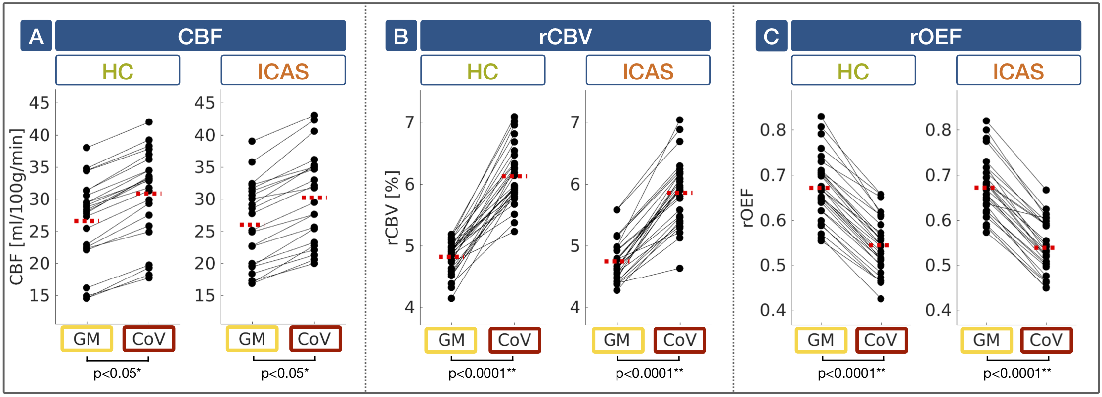
Figure 4: Hemodynamic characteristics in high-CoV voxels. CBF (A), rCBV (B) and rOEF (C) are compared in grey matter (GM; yellow) vs. high-CoV voxels (CoV; red) in healthy
controls (HC; green) and ICAS patients (orange). Dots show mean parameter
values, black lines connect the same subject’s mean values and red dashed lines
represent group average values. CBF was systematically lower due to background suppression13
and rOEF elevated due to T2 bias26. In high-CoV voxels of both groups, all parameters showed significant effects. While CBF (A) and rCBV (B) were higher, rOEF was
lower (C).
