Manali Balasaheb Jadhav1, Richa Singh Chauhan2, Priyanka Tupe Waghmare3, Archit Rajan4, Abhilasha Indoria2, Jitender Saini5, Vani Santosh6, Madhura Ingalhalikar4, and Subhas Konar7
1Symbiosis Center for Medical Image Analysis, Pune, India, 2Radiology, National Institute of Mental Health and Neuroscieces, Bengaluru, India, 3Symbiosis Institute on Technology, Pune, India, 4Symbiosis Centre for Medical Image Analysis, Pune, India, 5Radiology, National Institute of Mental Health and Neuroscieces, Pune, India, 6Neuropathology, National Institute of Mental Health and Neuroscieces, Bengaluru, India, 7Neurosurgery, National Institute of Mental Health and Neuroscieces, Bengaluru, India
1Symbiosis Center for Medical Image Analysis, Pune, India, 2Radiology, National Institute of Mental Health and Neuroscieces, Bengaluru, India, 3Symbiosis Institute on Technology, Pune, India, 4Symbiosis Centre for Medical Image Analysis, Pune, India, 5Radiology, National Institute of Mental Health and Neuroscieces, Pune, India, 6Neuropathology, National Institute of Mental Health and Neuroscieces, Bengaluru, India, 7Neurosurgery, National Institute of Mental Health and Neuroscieces, Bengaluru, India
Our random forest classifier with radiomics
computed from multi-modal MRI provides high discriminative accuracy in
predicting H3K27M mutation in midline glioma.
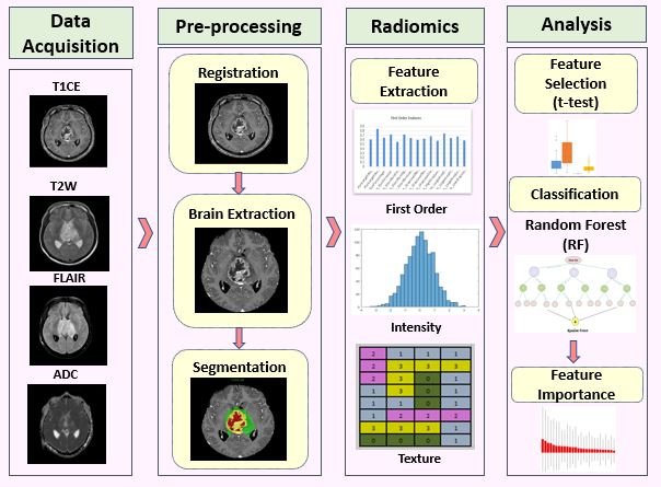
Figure 1:
Processing pipeline for radiomics analysis and classification for histone
H3K27M mutation
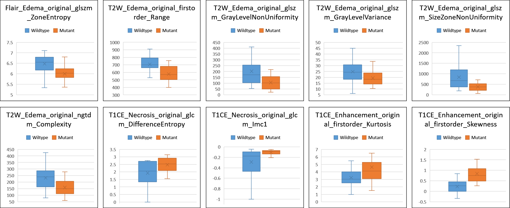
Figure 4: Box plot for top 10 most important features obtained using random forest classifier