Kiarash Ghassaban1,2, Chao Chai3, Huiying Wang4, Tong Zhang3, Jinxia Zhu5, Xianchang Zhang5, E. Mark Haacke1,2, and Shuang Xia3
1Department of Radiology, Wayne State University, Detroit, MI, United States, 2SpinTech, Inc., Bingham Farms, MI, United States, 3Department of Radiology, Tianjin First Central Hospital, Tianjin, China, 4Department of Neurology, Tianjin Medical University General Hospital Airport Site, Tianjin, China, 5MR Collaboration, Siemens Healthcare Ltd, Beijing, China
1Department of Radiology, Wayne State University, Detroit, MI, United States, 2SpinTech, Inc., Bingham Farms, MI, United States, 3Department of Radiology, Tianjin First Central Hospital, Tianjin, China, 4Department of Neurology, Tianjin Medical University General Hospital Airport Site, Tianjin, China, 5MR Collaboration, Siemens Healthcare Ltd, Beijing, China
The iRBD patients
had a higher incidence of Nigrosome-1 loss and increased iron in the right
dentate nucleus. Cognitive and motor impairment scores were associated with
iron in some of the deep gray matter structures.
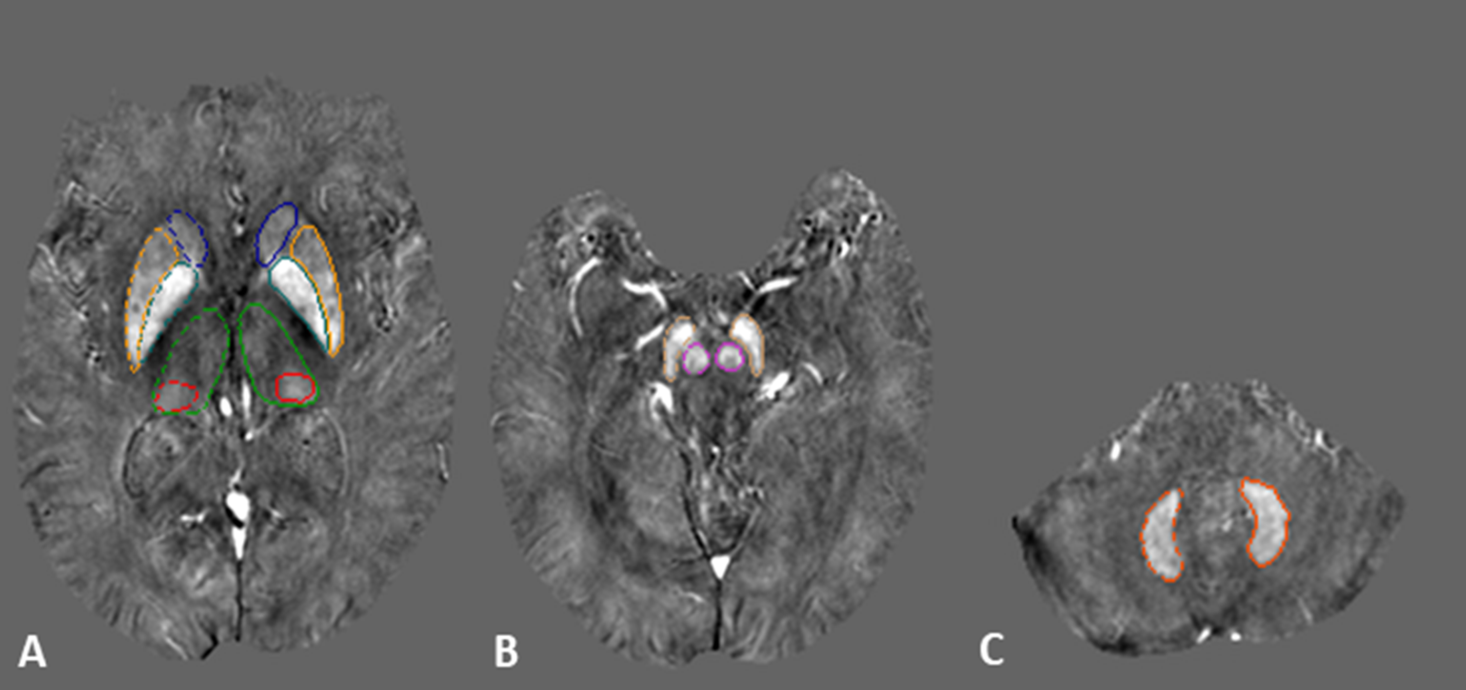
Figure
1. Regions of interest traced on quantitative
susceptibility maps on three representative slices showing deep gray matter
nuclei in A) the basal ganglia; blue: caudate nucleus, orange: putamen, cyan:
globus pallidus, green: thalamus, red: pulvinar thalamus. B) midbrain; purple:
red nucleus, yellow: substantia nigra. C) cerebellum; orange: dentate nucleus.
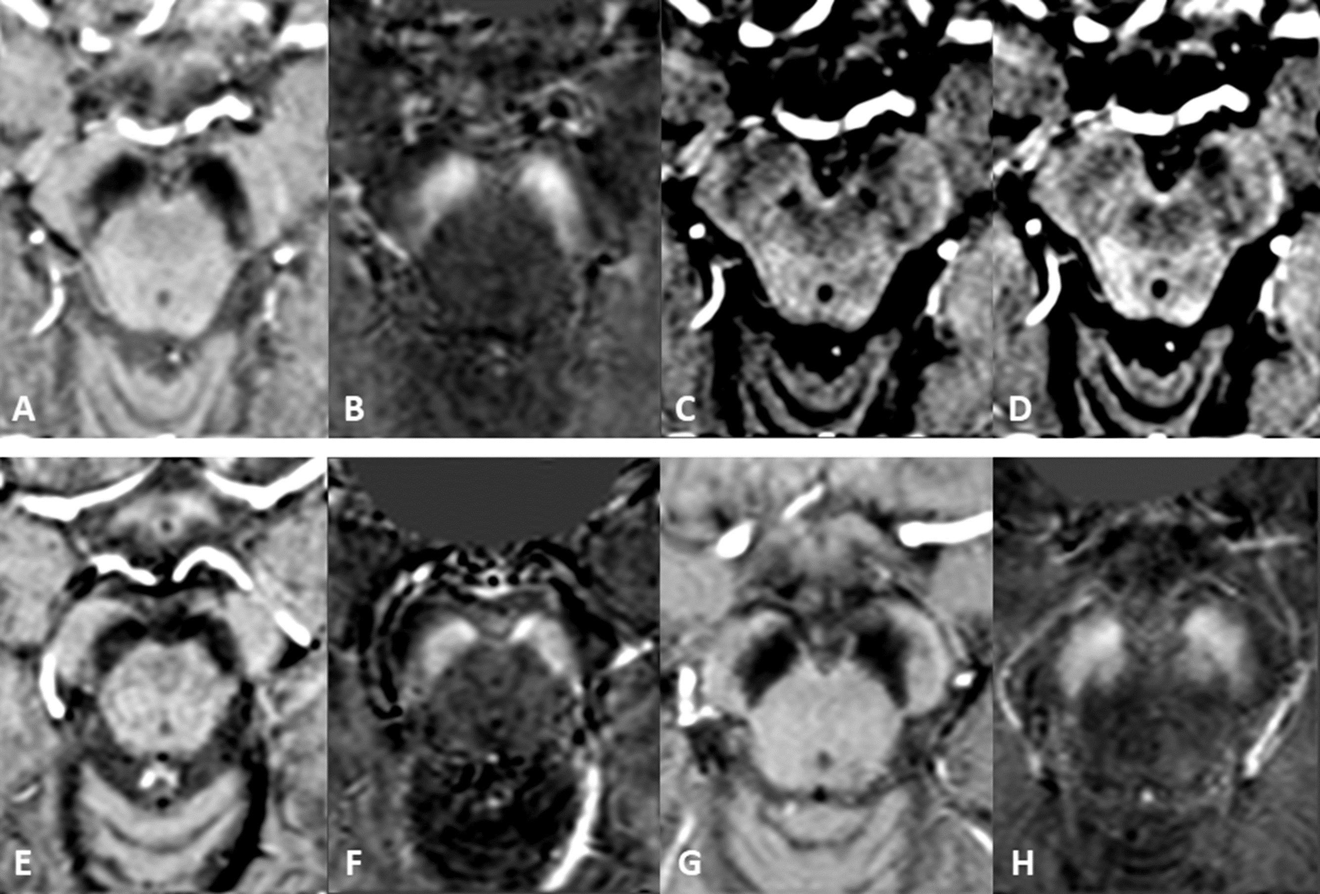
Figure 2. Nigrosome-1 sign in the substantia nigra. The top
row shows bilateral presence of N1 in A) tSWI, B) QSM, C) SWI and D) T2*W of a
healthy control. E, F) tSWI and QSM images of an iRBD patient with unilateral
loss of N1 in the left hemisphere. G, H) tSWI and QSM images of an iRBD patient
with bilateral loss of N1.