Baptiste MOREL1,2, Anne Sophie Piegay1, Maximilien Perivier1, Sandra Obry1, Bénédicte Maréchal3,4,5, Gian Franco Piredda3,4,5, Tom Hilbert3,4,5, Tobias Kober3,4,5, Clovis Tauber6, Pierre Castelnau6, and Jean Philippe Cottier1
1CHU de Tours, Tours, France, 2UMR 1253, iBrain, Université de Tours, INSERM, Tours, France, 3Advanced Clinical Imaging Technology, Siemens Healthcare AG, Lausanne, Switzerland, 4Department of Radiology, Lausanne University Hospital and University of Lausanne, Lausanne, Switzerland, 5LTS5, École Polytechnique Fédérale de Lausanne (EPFL), Lausanne, Switzerland, 6INSERM U1253, Tours, France
1CHU de Tours, Tours, France, 2UMR 1253, iBrain, Université de Tours, INSERM, Tours, France, 3Advanced Clinical Imaging Technology, Siemens Healthcare AG, Lausanne, Switzerland, 4Department of Radiology, Lausanne University Hospital and University of Lausanne, Lausanne, Switzerland, 5LTS5, École Polytechnique Fédérale de Lausanne (EPFL), Lausanne, Switzerland, 6INSERM U1253, Tours, France
A postprocessing brain MRI allowed obtaining automatically both volumetry
and T1 relaxometry values. It helps radiologists to
quantify brain abnormalities undetected in brain MRI in more than 80% of the initial exploration of children
with focal epilepsy of unknown cause.
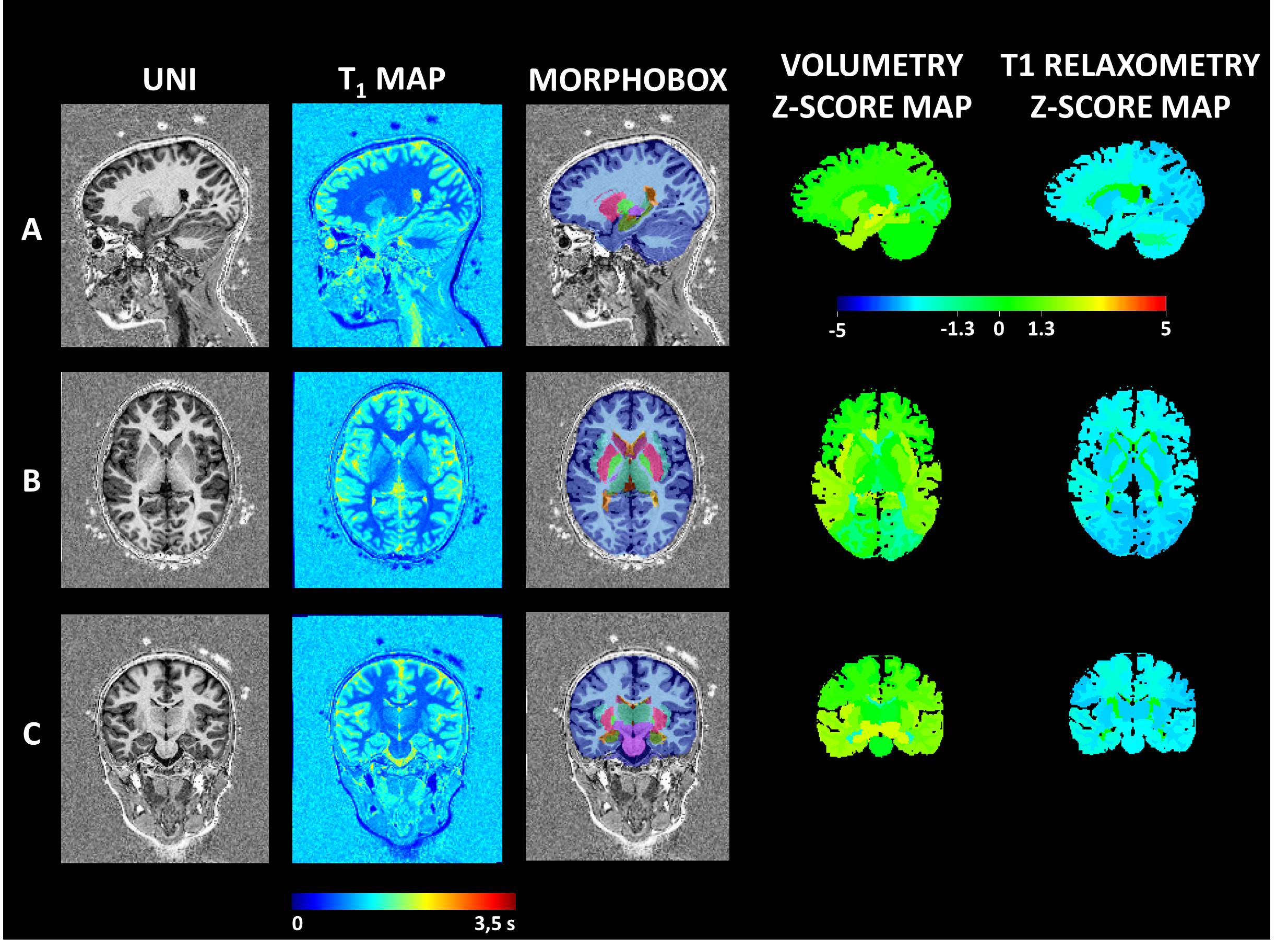
Sagittal, axial
and coronal views (line A, B and C) of the MP2RAGE sequence with the
postprocessing morphometric analysis results of a 6 years girl having left
focal epilepsia. UNI column is a highly contrasted T1 image. T1 relaxometry map
is the second column. Morphobox column represents the brain segmentation
obtained. A Volumetry and T1 relaxometry Z-score maps are also provided for a
visual analysis. We noticed a decreased T1 relaxometry values in the left
cortical occipital grey matter.