Humberto Monsivais1, Grace Francis2, Sandy Snyder1, Jonathan Kuhn3, and Ulrike Dydak1,4
1School of Health Sciences, Purdue University, West Lafayette, IN, United States, 2Department of Physics and Astronomy, Purdue University, West Lafayette, IN, United States, 3Mathematics and Statistics, Purdue University Northwest, Westville, IN, United States, 4Radiology and Imaging Scienes, Indiana University School of Medicine, Indianapolis, IN, United States
1School of Health Sciences, Purdue University, West Lafayette, IN, United States, 2Department of Physics and Astronomy, Purdue University, West Lafayette, IN, United States, 3Mathematics and Statistics, Purdue University Northwest, Westville, IN, United States, 4Radiology and Imaging Scienes, Indiana University School of Medicine, Indianapolis, IN, United States
Our results seem to reinforce the hypothesis that R1 is more sensitive and specific to brain Mn accumulation than PI, which indicates that currently R1 may be the best MRI marker to diagnose manganese induced neurotoxicity due to excess Mn accumulation in the brain.
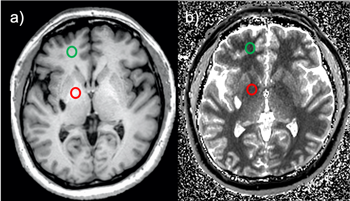
Figure 1. A) High-resolution T1-weighted image with circular ROIs in the globus pallidus (red) and frontal white matter (green) and visibly hyperintensities of the globus pallidus caused by Mn deposition. Similarly, b) T1-map, from which R1=1/T1 was calculated for several brain regions.
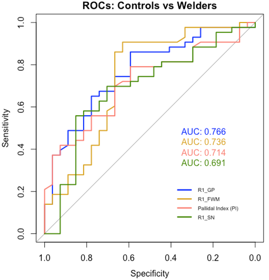
Figure 2. Receiver operating curves (ROCs) for PI, R1 in GP, FWM, and SN to distinguish between occupationally exposed subjects from unexposed controls. R1 in the GP yields the highest AUC (0.77) but only 86% sensitivity and 59% specificity. R1 in the FWM allows for similar discrimination between welders and controls with 91% sensitivity and 63% specificity (AUC = 0.74).