Ya-Jun Ma1, Catherine E Johnson2, Jonathan Wong1,3, Hyungseok Jang1, Roland Lee1, Eric Y Chang1,3, Zezong Gu2, and Jiang Du1
1UC San Diego, San Diego, CA, United States, 2Missouri University of Science and Technology, Rolla, MO, United States, 3VA Health System, San Diego, CA, United States
1UC San Diego, San Diego, CA, United States, 2Missouri University of Science and Technology, Rolla, MO, United States, 3VA Health System, San Diego, CA, United States
The 3D IR-UTE sequence allows direct
imaging of myelin and quantitative assessment of myelin density in mouse brain.
Melin loss in white matter of the brain induced by the open-field low intensity
blast can be reliably measured with the 3D IR-UTE sequence.

Figure
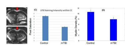
4. 3D IR-UTE imaging of myelin in a control mouse
(A) and a mouse four days after open-field LIB injury (B). LFB for the control
and mTBI mice shows a significant reduction in staining intensity within the
corpus callosum (CC) (red arrows) for the mTBI mouse (C), consistent with
demyelination induced by LIB. UTE-measured myelin density for the CC was
reduced by ~25% from 10.6±0.8% for the control mouse to 7.9±0.9% for the mTBI
mouse (F), largely consistent with LFB staining (C).
