Laura Biagi1,2, Rosa Pasquariello1, Raffaello Canapicchi 1, Chiara Ticci3, Claudia Dosi3, Graziella Donatelli2,4, Mauro Costagli1,2, Mirco Cosottini2,4,5, Roberta Battini3,6, Michela Tosetti1,2, and Network IDEA7
1Laboratory of Medical Physics and Magnetic Resonance, IRCCS Fondazione Stella Maris, Pisa, Italy, 2Imago7 Research Foundation, Pisa, Italy, 3Department of Developmental Neuroscience, IRCCS Fondazione Stella Maris, Pisa, Italy, 4Neuroradiology Unit, Azienda Ospedaliero-Universitaria Pisana, Pisa, Italy, 5Department of Translational Research and New Technologies in Medicine and Surgery, University of Pisa, Pisa, Italy, 6Department of Clinical and Experimental Medicine, University of Pisa, Pisa, Italy, 7Italian DEvelopmental Age Health Network (IDEA Network), Rome, Italy
1Laboratory of Medical Physics and Magnetic Resonance, IRCCS Fondazione Stella Maris, Pisa, Italy, 2Imago7 Research Foundation, Pisa, Italy, 3Department of Developmental Neuroscience, IRCCS Fondazione Stella Maris, Pisa, Italy, 4Neuroradiology Unit, Azienda Ospedaliero-Universitaria Pisana, Pisa, Italy, 5Department of Translational Research and New Technologies in Medicine and Surgery, University of Pisa, Pisa, Italy, 6Department of Clinical and Experimental Medicine, University of Pisa, Pisa, Italy, 7Italian DEvelopmental Age Health Network (IDEA Network), Rome, Italy
In a pediatric case of radio-chemotherapy induced leukoencephalopathy, 7T high-spatial resolution images are able to prove that most lesions radio-induced and/or dependent on combined chemo-radio treatment have an intracortical distribution.
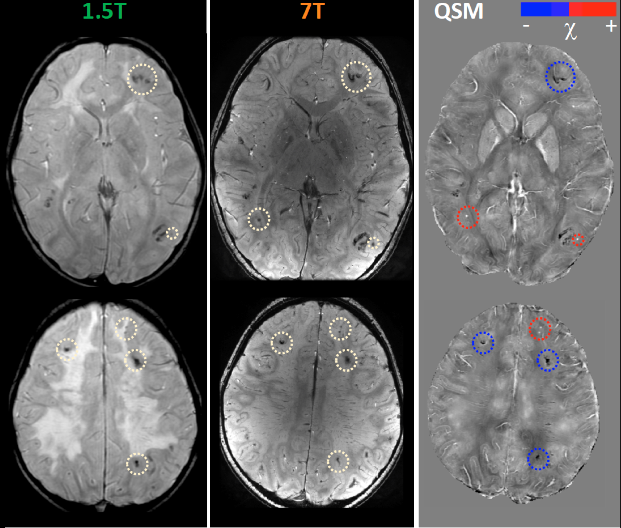
Figure 1: Comparison of 3D Gradient-Recalled Multi-Echo (SWAN) sequences at 1.5T (left column), 7T (central column), and QSM at 7T (right column). Hypo-intense signals in SWAN sequences (yellow circles) can be differentiated in paramagnetic formations (magnetic susceptibility >0, red circles), associable to vascular malformations, or mineralizing microangiopathy (magnetic susceptibility >0, blue circles).
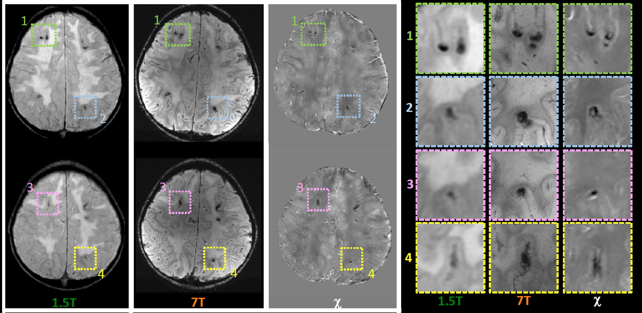
Figure 2: Examples of lesion representation at 1.5T (first column), 7T (second column), and QSM at 7T (third column). For each lesion surrounded by a colored box, the fourth column reports the corresponding zoomed depictions Whilst at 1.5T images their localization seems to be at the corticomedullary junction, 7T proves, thanks to its spatial resolution, that most lesions have an intracortical distribution.