wenjing huang1, jing zhang2, wanjun hu2, guangyao liu2, yanli jiang2, and shaoyu wang3
1Second Clinical School, Lanzhou University, Lanzhou, China, 2Lanzhou University Second Hospital, Lanzhou, China, 3Siemens Healthineers, Shanghai, China
1Second Clinical School, Lanzhou University, Lanzhou, China, 2Lanzhou University Second Hospital, Lanzhou, China, 3Siemens Healthineers, Shanghai, China
Our study demonstrated alterations
of the GMV and resting state FC in left parahippocampal gyrus, which may serve
as a biomarker for improving the understanding of cognitive decline and
depressive symptoms for mTBI in the acute setting.
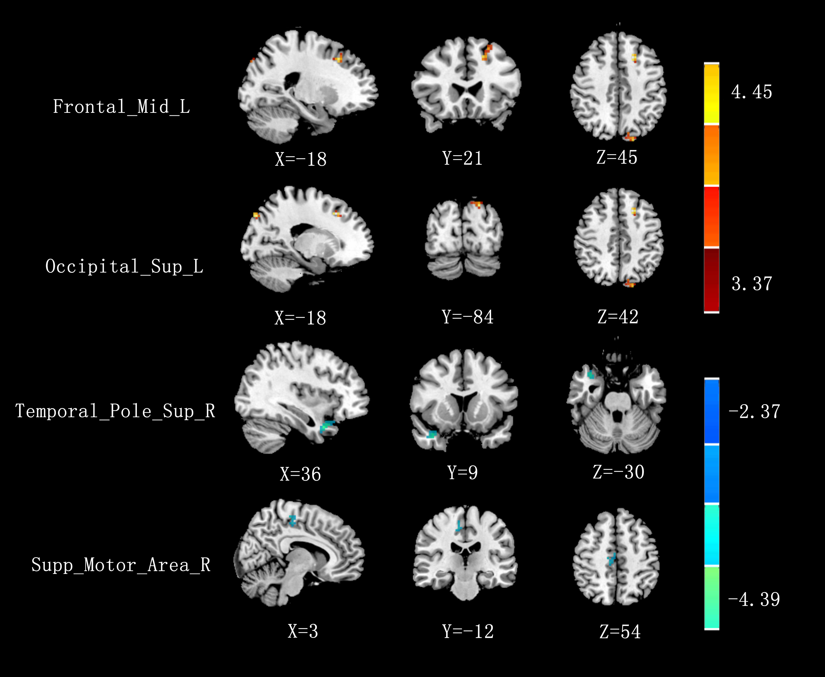
Fig 1 Significant differences in resting-state
FC between mTBI patients and healthy controls. The seed was located in the left
parahippocampal gyrus and the cold/warm color showed the regions that have
lower/higher correlation with seed point.
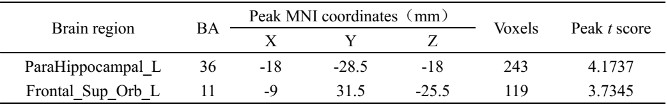
Table
1 GMV changes between mTBI patients and healthy controls. Multiple comparison correction
was performed using a threshold (p < 0.001) of individual voxel and a
cluster level p < 0.05(FWE corrected)