Kiran Thapaliya1,2, Donald Staines1, Sonya Marshall-Gradisnik1, and Leighton Barnden1
1Griffith University, Gold Coast, Australia, 2Centre for Advanced Imaging, The University of Queensland, Brisbane, Australia
1Griffith University, Gold Coast, Australia, 2Centre for Advanced Imaging, The University of Queensland, Brisbane, Australia
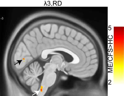
Brain stem abnormalities detected by diffusion tensor imaging could be the potential cause of Myalgic Encephalomyelitis/ Chronic fatigue syndrome.
We also found significant decrease of eigenvalues and mean diffusivity (MD) in brain stem of ME/CFS patients compared to healthy controls


