Linghan Kong1, Suhao Qiu1, Zhao He1, RunKe Wang1, Yu Chen1, Qiang He2, and Yuan Feng1
1Institute for Medical Imaging Technology, School of Biomedical Engineering, Shanghai Jiao Tong University, Shanghai, China, 2Shanghai United Imaging Healthcare Co Ltd, Shanghai, China
1Institute for Medical Imaging Technology, School of Biomedical Engineering, Shanghai Jiao Tong University, Shanghai, China, 2Shanghai United Imaging Healthcare Co Ltd, Shanghai, China
The cerebral blood flow
after applying vibration to the brain was measuring using 3D arterial spin
labeling. Results showed the vibration of brain at 30 Hz could reduce the
regional and global CBF.
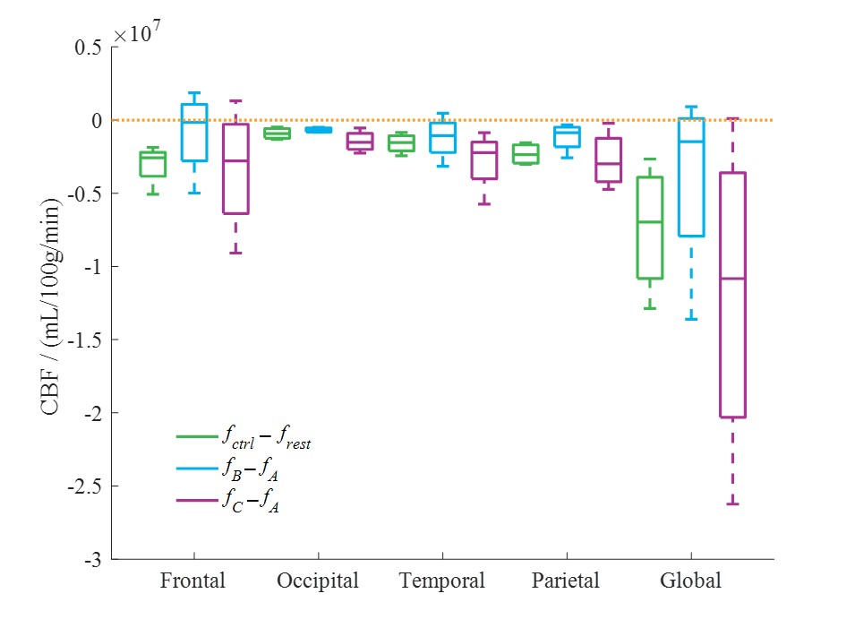
Figure 2: The
difference of CBF in each step. The CBF values were represented by f. Compared with resting state, the
regional and global mean CBF values of control state both decreased. Compared
with before vibration (fA),
the regional and global CBF values both decreased after two vibration (fB and fC). And compared with the first vibration, the CBF
decreased greater after the second vibration.
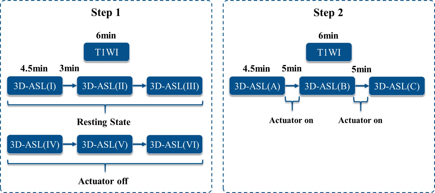
Figure
1: An illustration of the experimental workflow. In the first step, 3D-ASL was used to measure the CBF of subjects in the
resting state. Scan time was 4.5 minutes, and the scan interval was 3 minutes.
Then the subjects wear an actuator (off) to be scanned with 3D-ASL again
(Control). In
the second step, the subjects first wear an actuator (off) to be scanned
with 3D-ASL (A). And then the actuator was turned on for 5minutes. After the vibration, 3D-ASL
was used again to obtain the CBF of the subjects (B). To ensure
the stability of the results, the vibration and measurement was
repeated (C).