Anna Ivantsova1, Petr Menshchikov1,2,3, Andrei Manzhurtsev1,3, Maxim Ublinskii1,3, Alexey Yakovlev1,3,4, Ilya Melnikov1, Dmitrii Kupriyanov2, Tolib Akhadov1, and Natalia Semenova1,3,4
1Clinical and Research Institute of Emergency Paediatric Surgery and Traumatology, Moscow, Russian Federation, 2Clinical Science, LLC Philips Healthcare, Moscow, Russian Federation, 3Emanuel Institute of Biochemical Physics, Russian Academy of Sciences, Moscow, Russian Federation, 4Semenov Institute of Chemical Physics, Russian Academy of Sciences, Moscow, Russian Federation
1Clinical and Research Institute of Emergency Paediatric Surgery and Traumatology, Moscow, Russian Federation, 2Clinical Science, LLC Philips Healthcare, Moscow, Russian Federation, 3Emanuel Institute of Biochemical Physics, Russian Academy of Sciences, Moscow, Russian Federation, 4Semenov Institute of Chemical Physics, Russian Academy of Sciences, Moscow, Russian Federation
The main finding of the study is that the tNAA
signal reduction in WM after mTBI is associated with a decrease in the NAAG
concentration rather than a decrease in the NAA concentration, as was thought
previously.

Fig. 4. The NAAG (left) and NAA (right) MEGA-PRESS spectra
summed over the controls (blue) and patient (red) groups. The difference
between the spectra are shown in black. Summed NAAG signal (δ = 2.6 ppm)
significantly reduced in patient group as compared to controls.
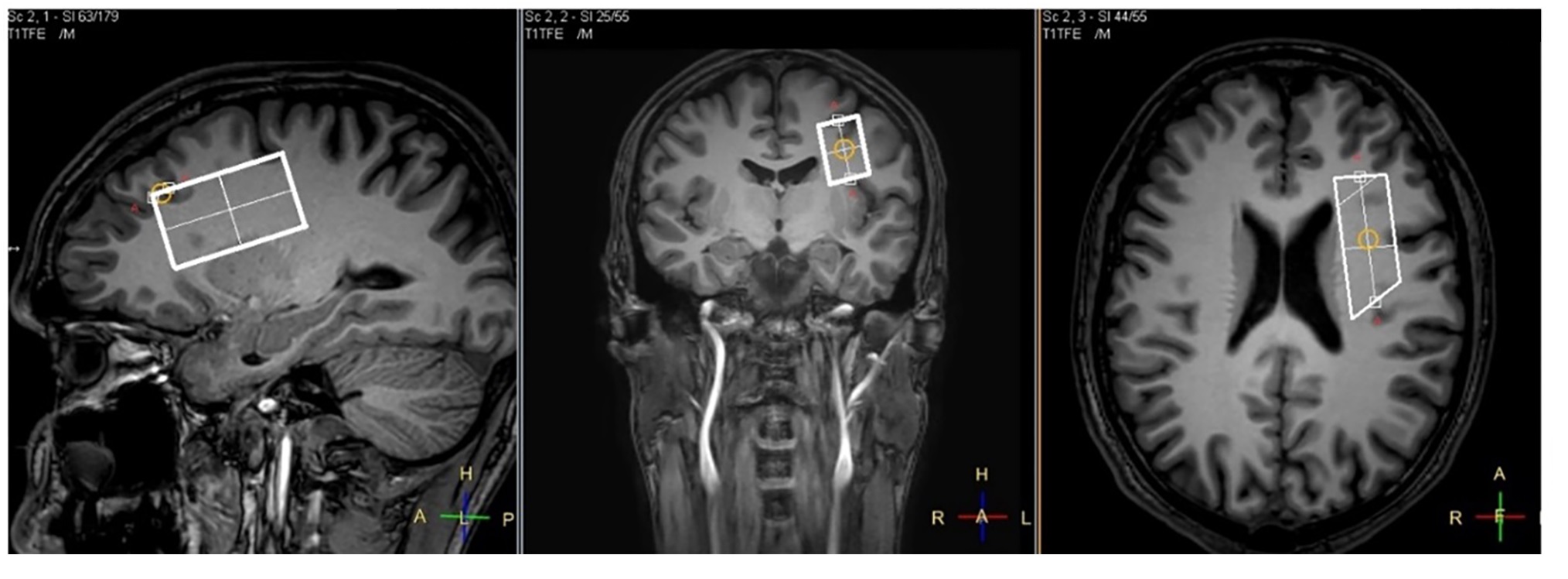
Fig. 1. Typical VOI localization: dorsolateral
pre-frontal area (WM-dominant brain region) VOI, 50×19×27 mm3