Mark David Platt1, Harish Poptani1, Antonius Plagge2, and Mahon Maguire1
1Molecular and Clinical Cancer Medicine, University of Liverpool, Liverpool, United Kingdom, 2Cellular and Molecular Physiology, University of Liverpool, Liverpool, United Kingdom
1Molecular and Clinical Cancer Medicine, University of Liverpool, Liverpool, United Kingdom, 2Cellular and Molecular Physiology, University of Liverpool, Liverpool, United Kingdom
In vivo
MRI demonstrated significant differences between the whole brain, cerebellar
and corpus callosum volumes of adult TRAPPC9 knockout mice and wildtype
controls. Behavioural assays also revealed significant differences in learning
ability.
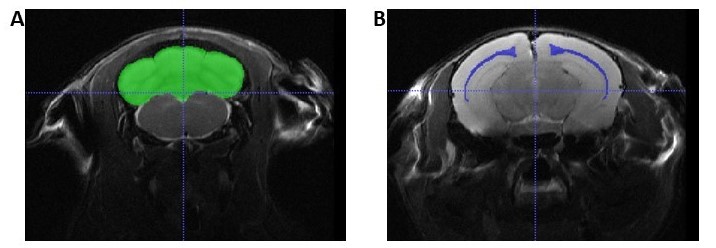
Figure 1: Example slices of the in vivo T2-weighted images with segmentation masks for the
cerebellum (A) and corpus callosum (B) overlaid in ITK-Snap (http://www.itksnap.org).
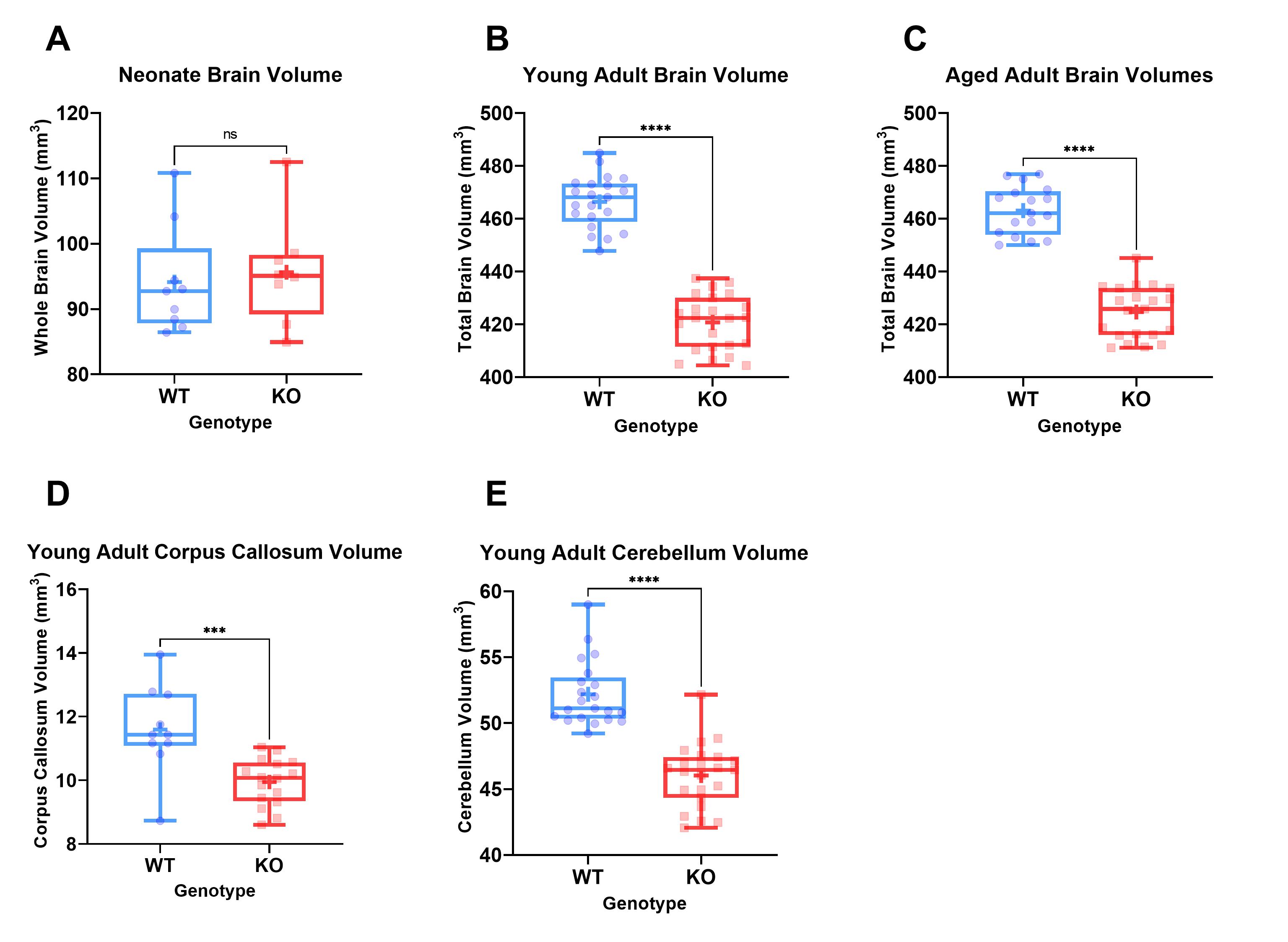
Figure 2: Boxplots showing the full range, median and
average (+) values for brain volumes in neonates (A), young adults (B), aged adults
(C) and regional volumes of corpus callosum (D) and cerebellum (E). Significance
levels are indicated by: ns = P > 0.05, * = P ≤ 0.05, **= P ≤ 0.01, ***= P ≤ 0.001, ****=
P ≤ 0.0001.