Vladimir Grouza1,2, Zhe Wu1,3, Marius Tuznik1,2, Hooman Bagheri4, Dan Wu5, Alan C Peterson2,4,6, and David Rudko1,2,7
1McConnell Brain Imaging Centre, Montreal Neurological Institute and Hospital, Montreal, QC, Canada, 2Department of Neurology and Neurosurgery, McGill University, Montreal, QC, Canada, 3Techna Institute, University Health Network, Toronto, ON, Canada, 4Department of Human Genetics, McGill University, Montreal, QC, Canada, 5Russell H. Morgan Department of Radiology & Radiological Science, Johns Hopkins University, Baltimore, MD, United States, 6Gerald Bronfman Department of Oncology, McGill University, Montreal, QC, Canada, 7Department of Biomedical Engineering, McGill University, Montreal, QC, Canada
1McConnell Brain Imaging Centre, Montreal Neurological Institute and Hospital, Montreal, QC, Canada, 2Department of Neurology and Neurosurgery, McGill University, Montreal, QC, Canada, 3Techna Institute, University Health Network, Toronto, ON, Canada, 4Department of Human Genetics, McGill University, Montreal, QC, Canada, 5Russell H. Morgan Department of Radiology & Radiological Science, Johns Hopkins University, Baltimore, MD, United States, 6Gerald Bronfman Department of Oncology, McGill University, Montreal, QC, Canada, 7Department of Biomedical Engineering, McGill University, Montreal, QC, Canada
The application of BSS-rPCA MWF estimation was evaluated with the aide of a well characterized hypomyelinating mouse model. Resultant estimates were found to correlate well with Mbp/Golli mRNA expression levels.
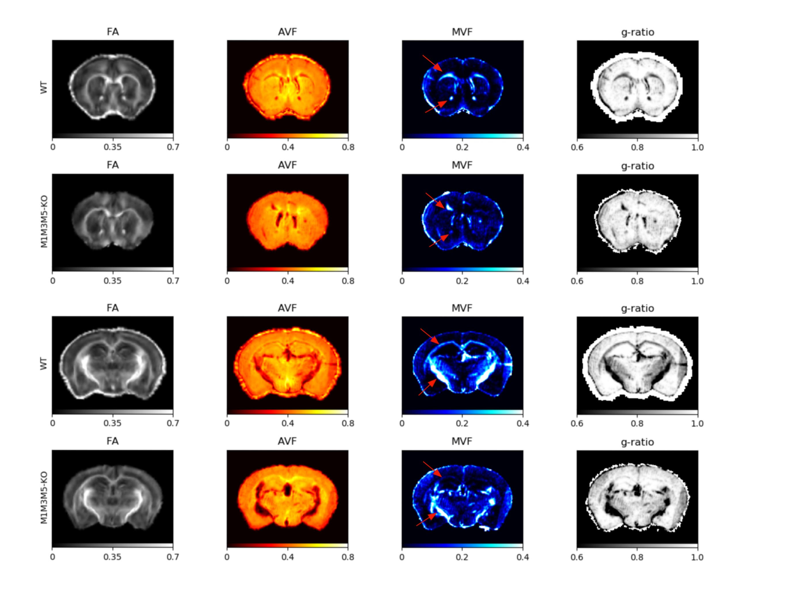
Figure 1: Coronal slices showing FA, AVF, MVF, and g-ratio parameter maps in WT
and M1M3M5KO mice. Selected slices in the top two rows permit
visualization of the genu of the corpus callosum and
the anterior commissure white matter tract. The bottom two rows display
the internal capsule and
the splenium of the corpus callosum in the mouse brain. The difference
myelin volume
fraction signal between WT and M1M3M5KO is evident based on red arrows
overlaid on the mouse brain images.

Associations between myelin volume fraction, derived from
BSS rPCA-based multi-component T2* analysis, and relative myelin basic protein mRNA levels in three major white matter tracts of the mouse brain. Mbp mRNA levels were derived using qRT-PCR as described in Bagheri et al.1 In
anterior commissure, splenium of the corpus callosum and internal
capsule white matter, MVF scales linearly with relative Mbp mRNA. This
supports the conjecture that Mbp mRNA level is a critical determinant of MRI-sensitive
myelin bilayer properties.
