Xueru Liu1,2, Huilin Hong3, Zhentao Zuo1,2,4, Hui Zhao3, Rui Tian3, Yongqing Zhang2,3,4, and Yan Zhuo1,2,4
1State Key Laboratory of Brain and Cognitive Science, Institute of Biophysics, Chinese Academy of Sciences, BeiJing, China, 2University of Chinese Academy of Sciences, Beijing, China, 3State Key Laboratory of Molecular Developmental Biology, Institute of Genetics and Developmental Biology, Chinese Academy of Sciences, Beijing, China, 4CAS Center for Excellence in Brain Science and Intelligence Technology, Chinese Academy of Sciences, Beijing, China
1State Key Laboratory of Brain and Cognitive Science, Institute of Biophysics, Chinese Academy of Sciences, BeiJing, China, 2University of Chinese Academy of Sciences, Beijing, China, 3State Key Laboratory of Molecular Developmental Biology, Institute of Genetics and Developmental Biology, Chinese Academy of Sciences, Beijing, China, 4CAS Center for Excellence in Brain Science and Intelligence Technology, Chinese Academy of Sciences, Beijing, China
Socially deprived dogs strengthened brain function connectivity
within frontal cortex and weakened function connectivity between PFC with the
visual and auditory cortex. Diffusion metrics observed
demyelination in frontal, temporal, and insula regions after 4 weeks social
deprivation.
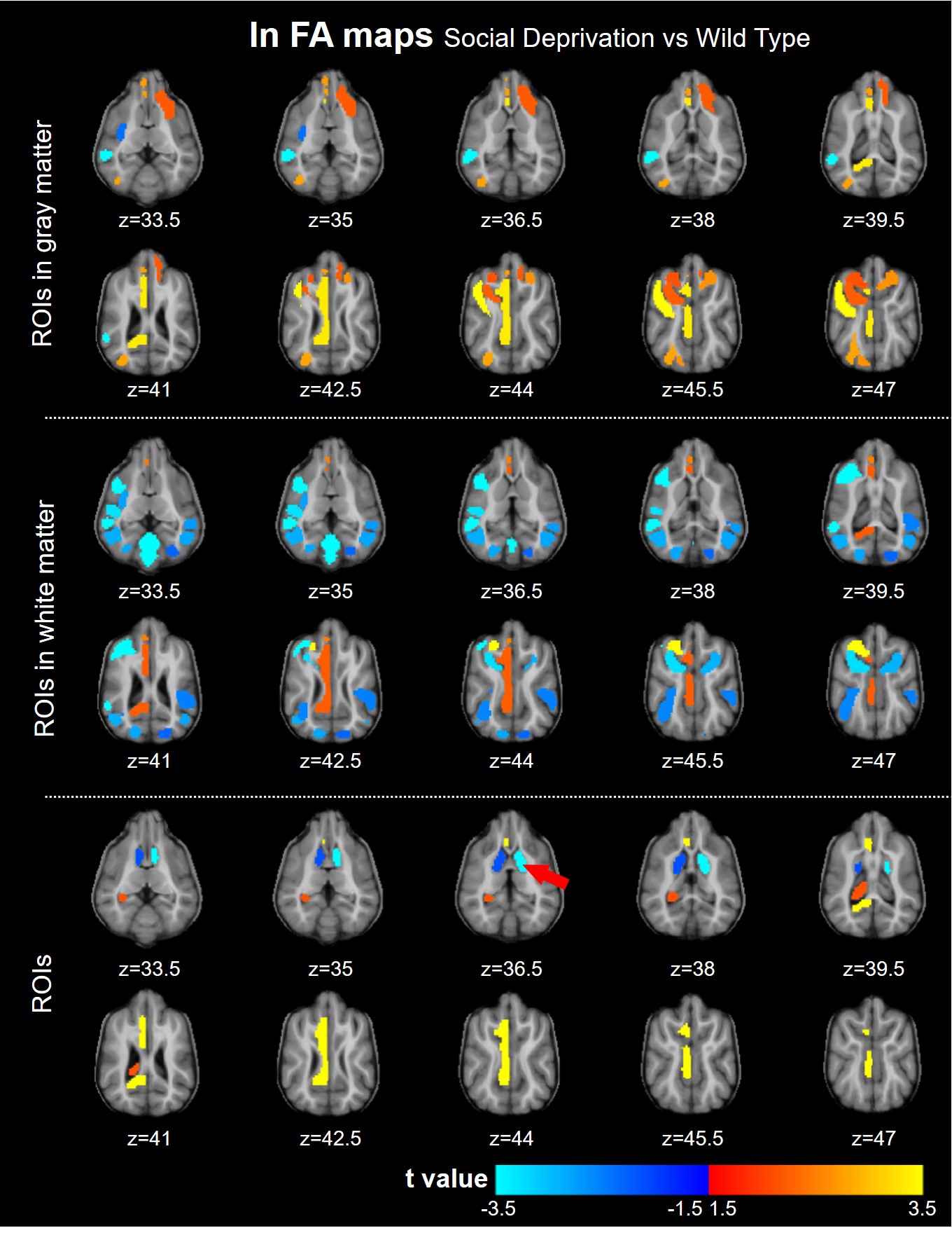
Figure 4. Statistics
of FA values of 86 ROIs in the cortex, white matter, and subcortical nucleus
between SD and WT groups. The t values
were obtained by unpaired t-test of
FA maps from two groups.
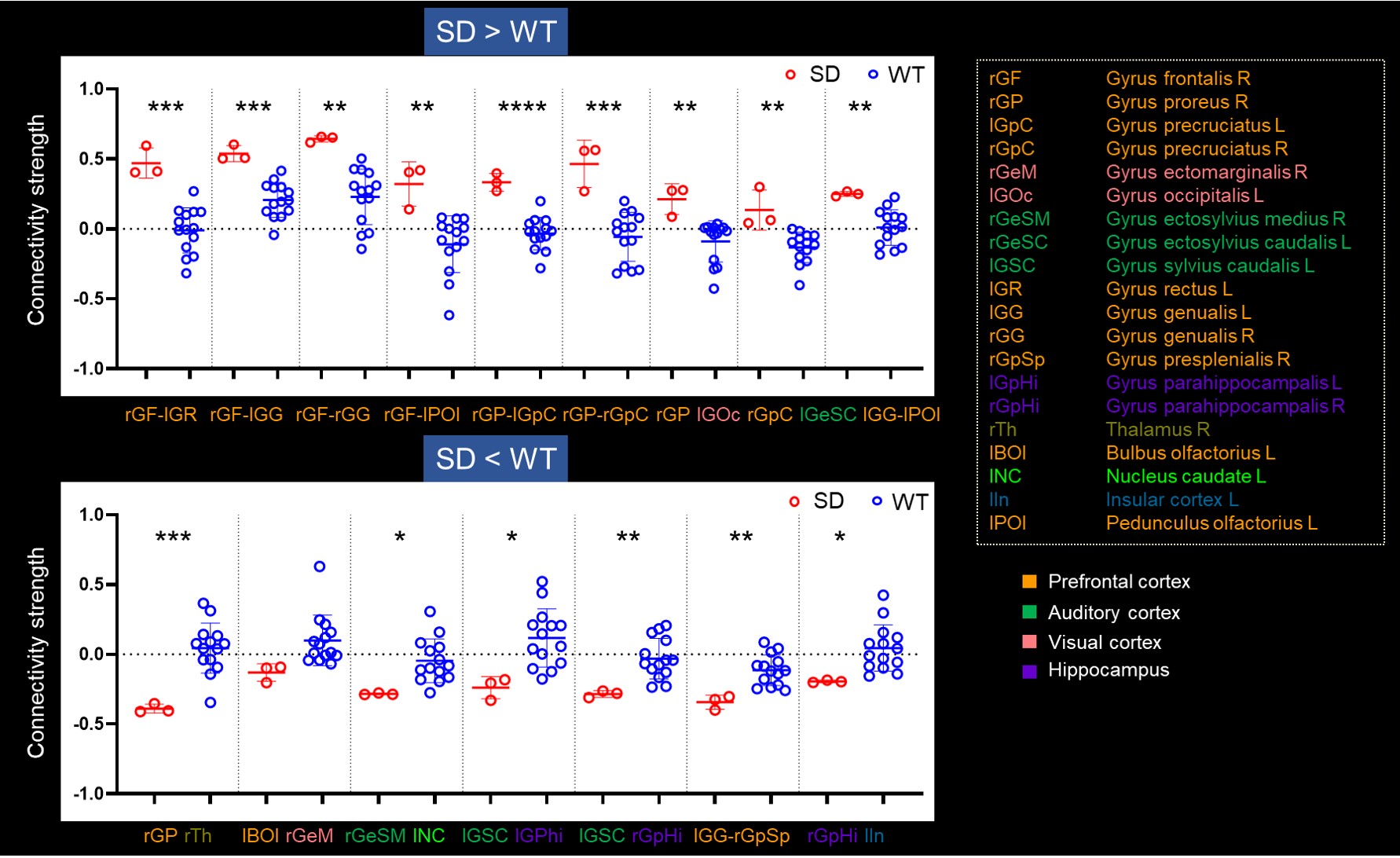
Figure 3. Comparison
of connectivity strength between social deprivation and wild type groups. The r
values of 86 ROIs were compared between SD and WT. The two charts showed ROIs
with significant differences. The red arrows indicated that the FA values
in the temporal lobe and nucleus caudatus of socially deprived were significantly
reduced compared with WT.