Zhixing Wang1, Xue Feng1, John Mugler2, and Craig Meyer1
1Biomedical Engineering, University of Virginia, Charlottesville, VA, United States, 2Radiology & Medical Imaging, University of Virginia, Charlottesville, VA, United States
1Biomedical Engineering, University of Virginia, Charlottesville, VA, United States, 2Radiology & Medical Imaging, University of Virginia, Charlottesville, VA, United States
This study describes a new approach to obtain T2-weighted and fluid-attenuated inversion recovery (FLAIR) images simultaneously in a short scan time using a spiral-ring Turbo Spin-echo sampling strategy combined with a 180°(y)-90°(x) driven-inversion preparation RF pulse.

Figure 1. Pulse sequence diagram showing the sampling strategy, which includes the time-multiplexed multislice scheme, SPRING TSE in/out variant data acquisition, and driven-inversion RF pulses. The boxes with stripes show slices for the T2-weighted acquisition, while the open boxes show slices for the FLAIR acquisition. The time interval between boxes with the same number is set to TI. A SPRING TSE in/out variant sampling scheme is used in each box for data acquisition, while a 180°-90° RF pair is applied at the end of the echo train for boxes with stripes to achieve driven inversion.
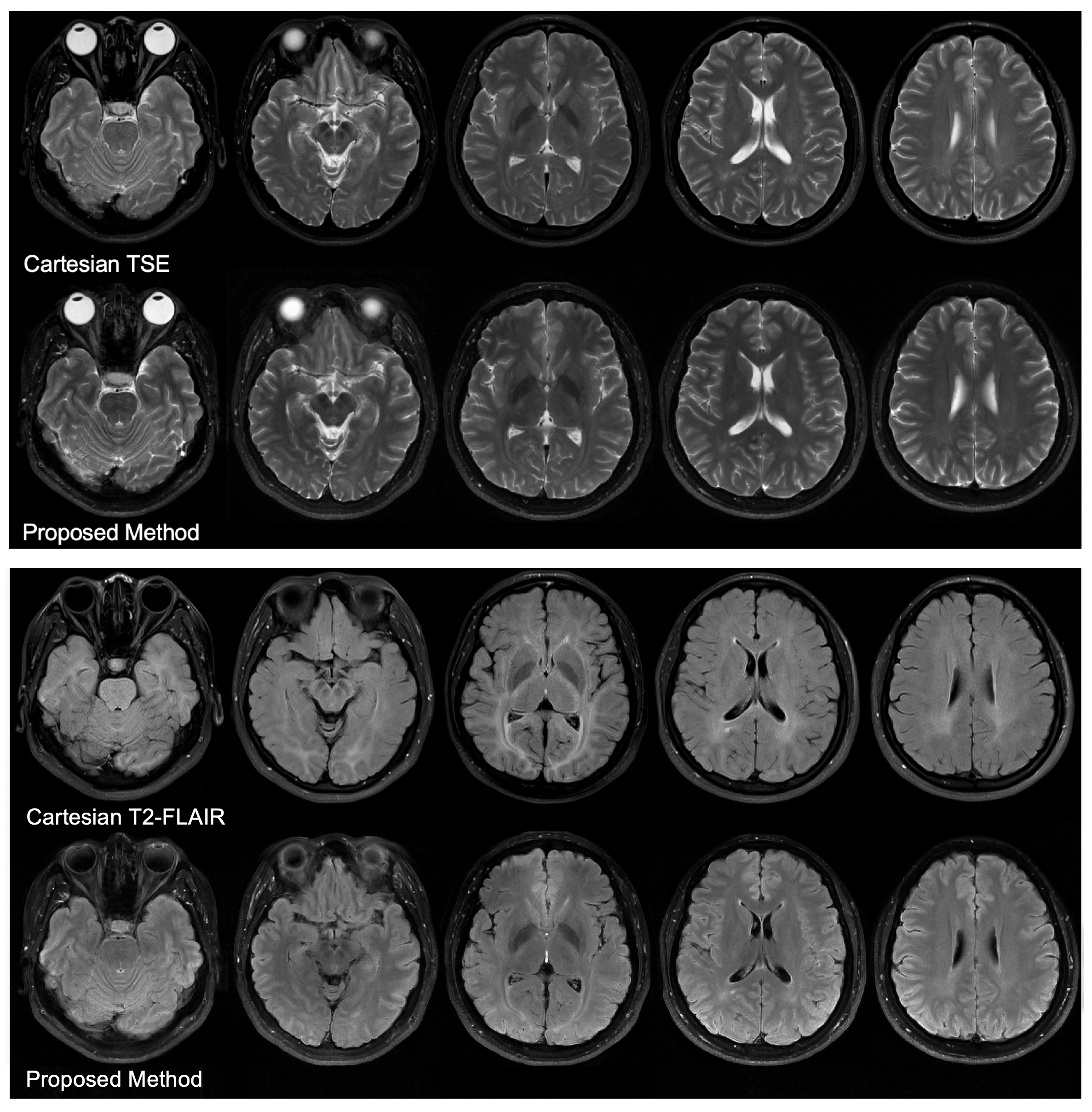
Figure 3. Comparison of in-vivo images acquired using standard Cartesian TSE for T2-weighted images, Cartesian FLAIR for fluid-attenuated images, and the proposed method for both the T2-weighted and fluid-attenuated images. Both the T2-weighted images (top panel) and FLAIR images (bottom panel) from the proposed method show image quality and contrast similar to the conventional images.