Tracy Ssali1, Lucas Narciso1,2, Justin Hicks1,2, Matthais Günther3, Frank Prato1,2, Udunna Anazodo1,2, Elizabeth Finger4, and Keith St Lawrence1,2
1Lawson Health Research Institute, London, ON, Canada, 2Department of Medical Biophysics, Western University, London, ON, Canada, 3Fraunhofer Institute for Medical Image Computing MEVIS, Bremen, Germany, 4Department of Clinical Neurological Sciences, Western University, London, ON, Canada
1Lawson Health Research Institute, London, ON, Canada, 2Department of Medical Biophysics, Western University, London, ON, Canada, 3Fraunhofer Institute for Medical Image Computing MEVIS, Bremen, Germany, 4Department of Clinical Neurological Sciences, Western University, London, ON, Canada
This work highlights the potential of ASL for identifying regional hypoperfusion in FTD patients. While 15O-water PET data showed greater sensitivity, similar areas of hypoperfusion were identified by ASL, particularly for relative CBF maps which reduced inter-subject variability.
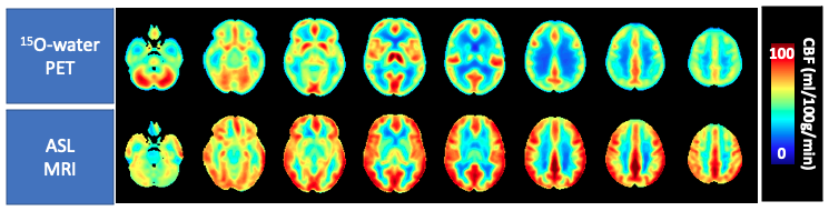
Figure 1: Mean control perfusion maps measured by ASL and 15O-water (whole brain CBF = 61.6 ± 12.4 ml/100g/min).
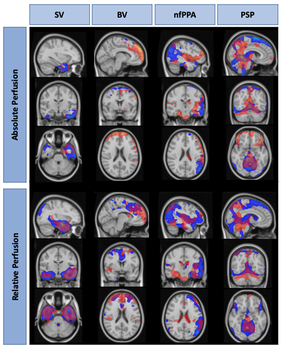
Figure 2: T-maps generated using relative and absolute CBF measured by ASL and 15O-water in 4 exemplary FTD patients. Areas in red (ASL) and blue (15O-water) indicate regions of significant hypoperfusion in the patient participant compared to the control group. sv = semantic variant, bv=behavioural variant, nfPPA = non-fluent primary progressive aphasia, psp = progressive supranuclear palsy.