Tales Santini1, Minseok Koo1, Nadim Farhat1, Vinicius P. Campos2, Salem Alkhateeb1, Marcelo A. C. Vieira2, Meryl A Butters1, Caterina Rosano1, Howard J Aizenstein1, Joseph Mettenburg1, Enrico M. Novelli1, and Tamer S Ibrahim1
1University of Pittsburgh, Pittsburgh, PA, United States, 2University of Sao Paulo, Sao Carlos, Brazil
1University of Pittsburgh, Pittsburgh, PA, United States, 2University of Sao Paulo, Sao Carlos, Brazil
We collected and processed 7T MRI images from individuals with sickle cell
disease and matched controls. Individuals with SCD have significantly smaller
volumes of the DG+CA2+CA3 hippocampal region. Other subregions also showed a
trend towards smaller volumes
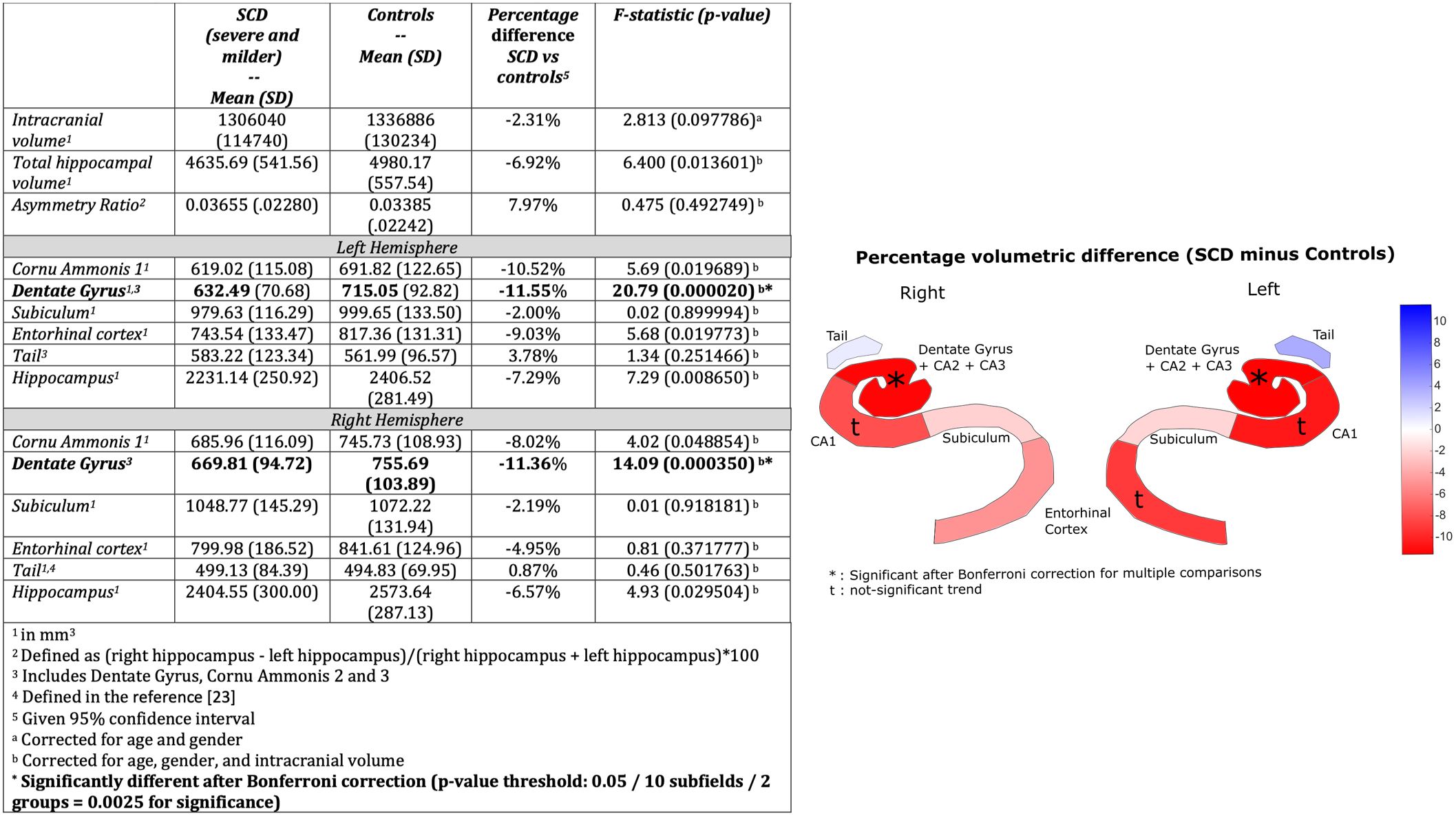
Neuroimaging characteristics. Mean volume (in mm3),
standard deviation and percent difference.
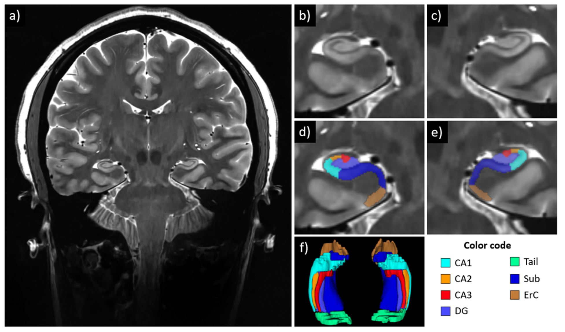
Example of hippocampal subfields segmentation in a subject
with SCD. a: coronal slice of the T2-weighted image, acquired at 7T with
resolution 0.375x0.375x1.5mm2; b,c: zoom-in images showing details
of the hippocampus structure, subject right and left, respectively; d,e:
hippocampus subfield segmentations overlaying the T2-weighted image, subject
right and left, respectively; f: 3D reconstruction of the hippocampal subfield
segmentations.