Thomas Troalen1, Arnaud Le Troteur2, Sylviane Confort-Gouny2, Patrick Vioux2, Claire Costes2, Lauriane Pini2, Jean-Philippe Ranjeva2, Maxime Guye2, and Ludovic de Rochefort2
1Siemens Healthcare SAS, Saint-Denis, France, 2CRMBM UMR7339 CNRS Aix-Marseille Université, Marseille, France
1Siemens Healthcare SAS, Saint-Denis, France, 2CRMBM UMR7339 CNRS Aix-Marseille Université, Marseille, France
This work demonstrates the ability to accelerate
multi gradient echo sequences for a joint R2* and QSM in the brain. Combining this sequence with a state-of-the art automatic
post-processing pipeline, we propose here a standardized whole brain clinical
protocol of 5 minutes.
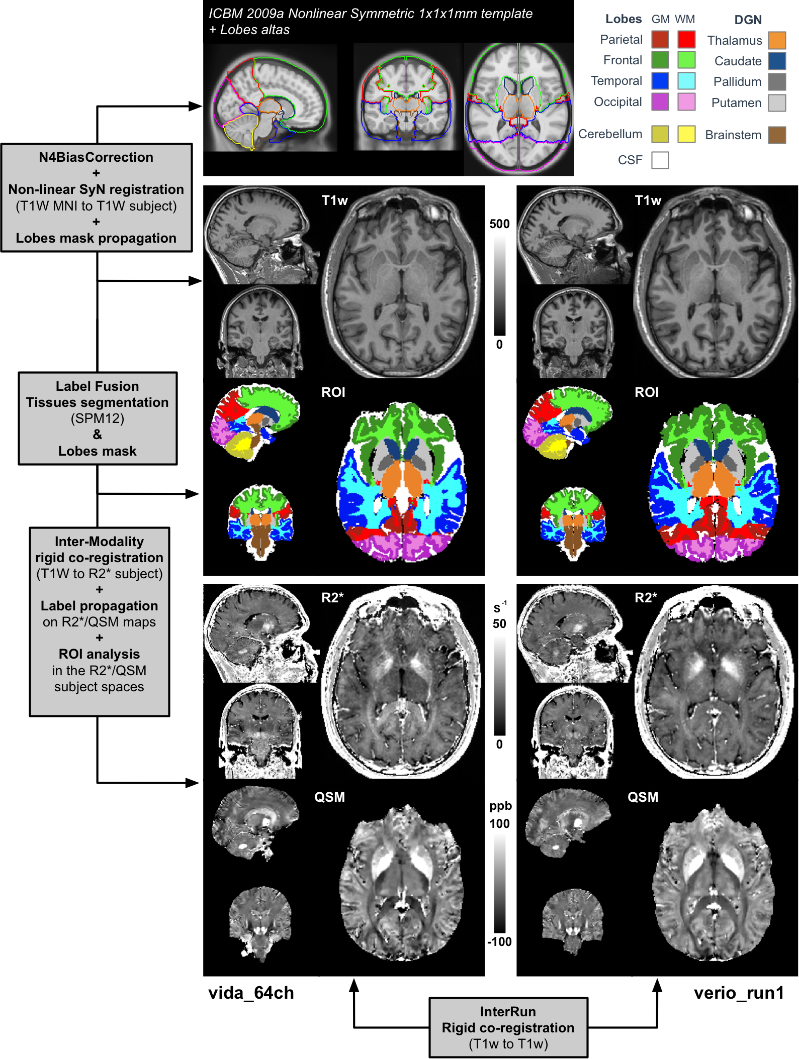
Figure 1: Automatic
processing pipeline: The ICBM 2009a nonlinear symmetric MNI template was non-linearly
registered to the subject’s T1w space (using Vida_64Ch as reference).
Inter/Intra-run
co-registrations were performed (T1wRef-to-T1w, as well as T1w-to-R2*). Tissue
segmentation was achieved using SPM12 software using the default brain
probability maps. Labels were propagated to R2*/QSM space and restricted to
WM/GM tissue types. WM/GM lobes and cerebellum were extracted, as well as four
deep grey nuclei and the brainstem prior ROI analysis on the quantitative maps.
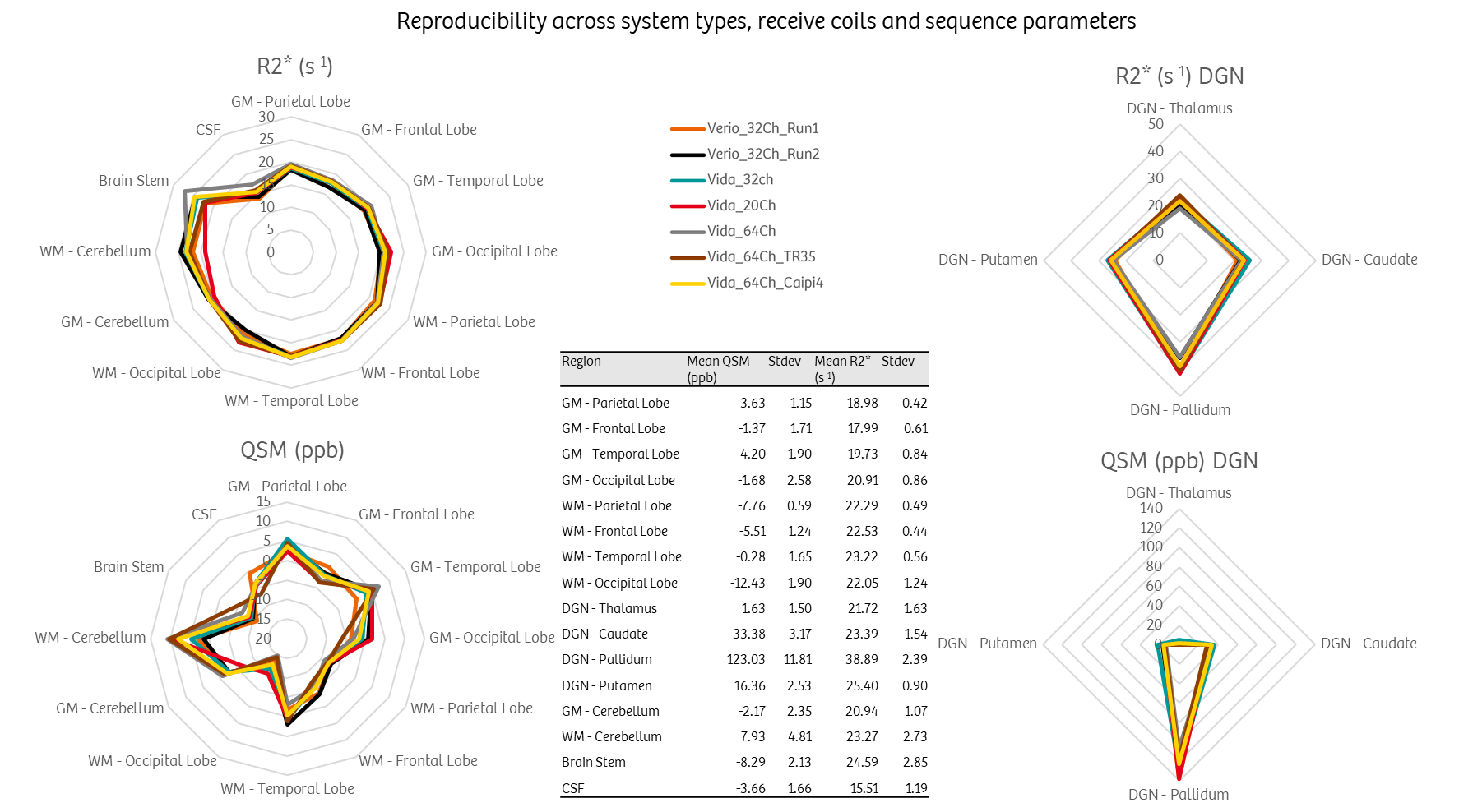
Reproducibility across
MR system types (Verio and Vida), receive head coils (20, 32 and 64 channels)
and sequence parameters. Note that the measured variability on the Vida with
different head coils was not different than the intra-run metric variations on
the Verio system. The signal drop caused by the increased acceleration factor
from 3 to 4 was not reflected in the R2* and QSM estimations, with values in
the same range as the other protocols. The central table reports mean and
standard deviation per segmented region across all measurements.