Jiaen Liu1, Peter van Gelderen1, Jacco A. de Zwart1, and Jeff H. Duyn1
1AMRI, LFMI, NINDS, National Institutes of Health, Bethesda, MD, United States
1AMRI, LFMI, NINDS, National Institutes of Health, Bethesda, MD, United States
High-resolution T2*-weighted 7 T MRI can be used to delineate intracortical structure owning to its high signal-to-noise and contrast-to-noise ratio. Reliable ultrahigh resolution T2*-weighted MRI can be reliably obtained using a navigator-based sequence with motion and B0 correction.
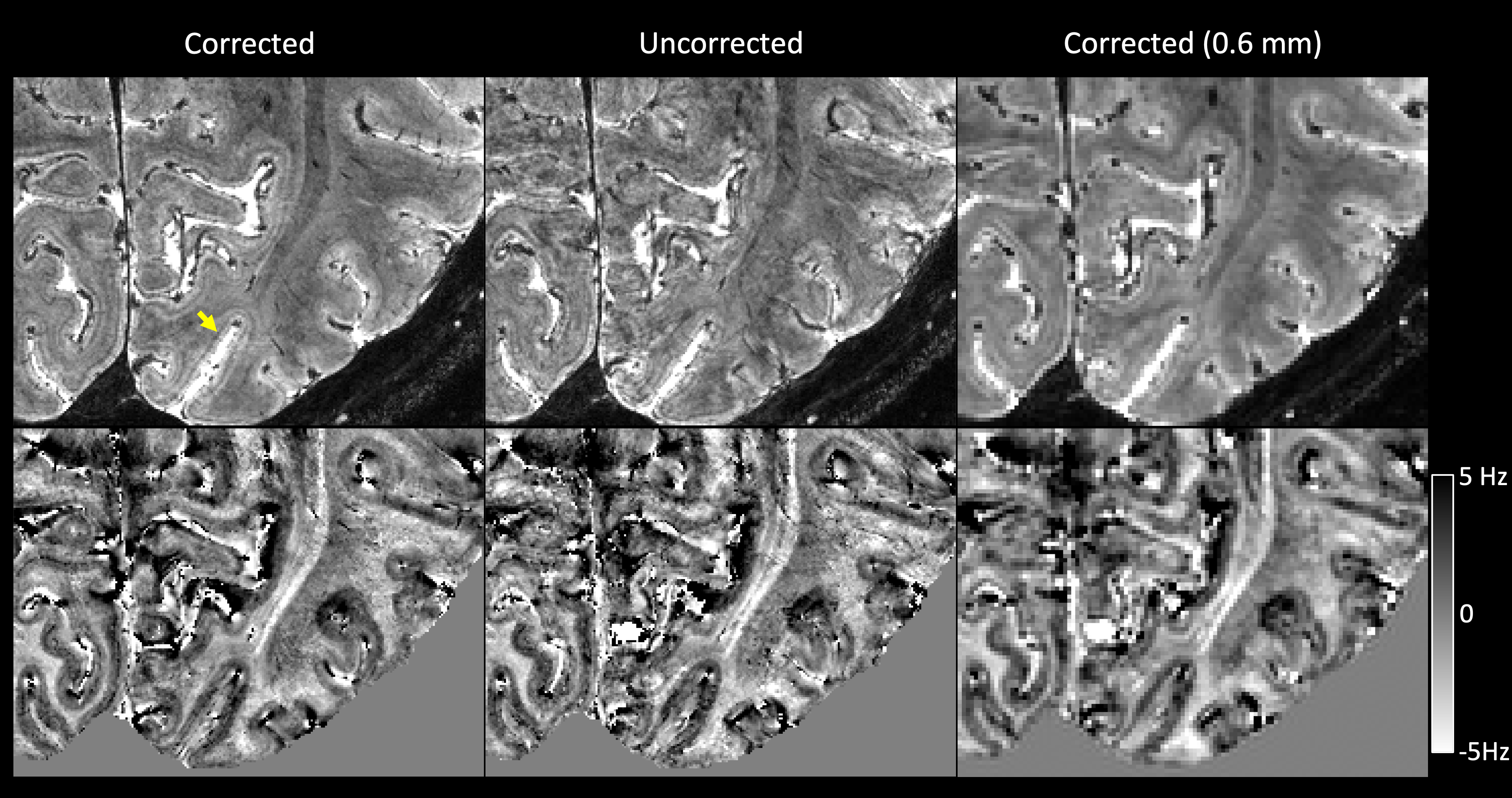
Fig. 1 Top row: magnitude of corrected, uncorrected and lower-resolution corrected (isotropic 0.6 mm) T2*-weighted MRI from one subject. The yellow arrow points to the hypointensive Line of Gennari in the primary visual cortex. Bottom row: the corresponding susceptibility-induced off-resonance frequency maps at 7 T. The Line of Gennari appears as a positive-frequency band (note darker equals more positive).
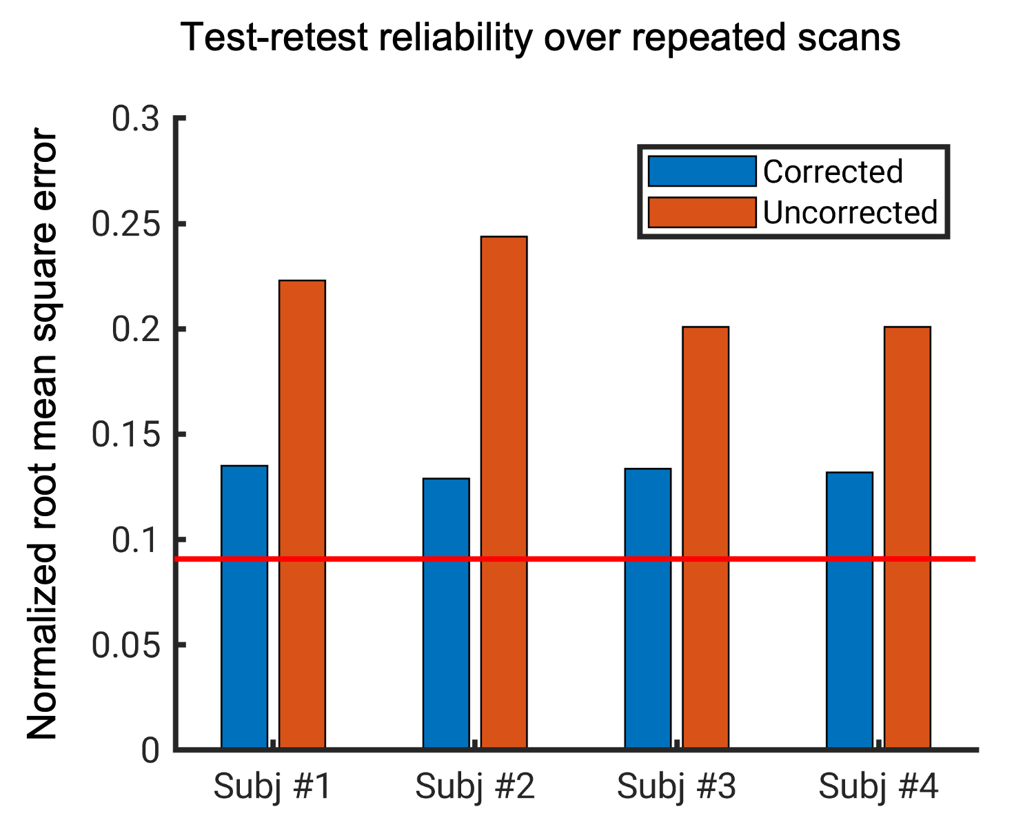
Fig. 2 Normalized root mean square error (NRMSE) between two repeated T2*-weighted scans for each subject. Red line represents the expected NRMSE caused by thermal noise.