Eric Seth Michael1, Franciszek Hennel1, and Klaas Paul Pruessmann1
1Institute for Biomedical Engineering, ETH Zurich and University of Zurich, Zurich, Switzerland
1Institute for Biomedical Engineering, ETH Zurich and University of Zurich, Zurich, Switzerland
The use of motion-compensated oscillating diffusion
gradients permitted high-resolution, interleaved acquisitions of the in-vivo
human brain. This implementation produced
images void of visible artifacts without the use of additional computational techniques.
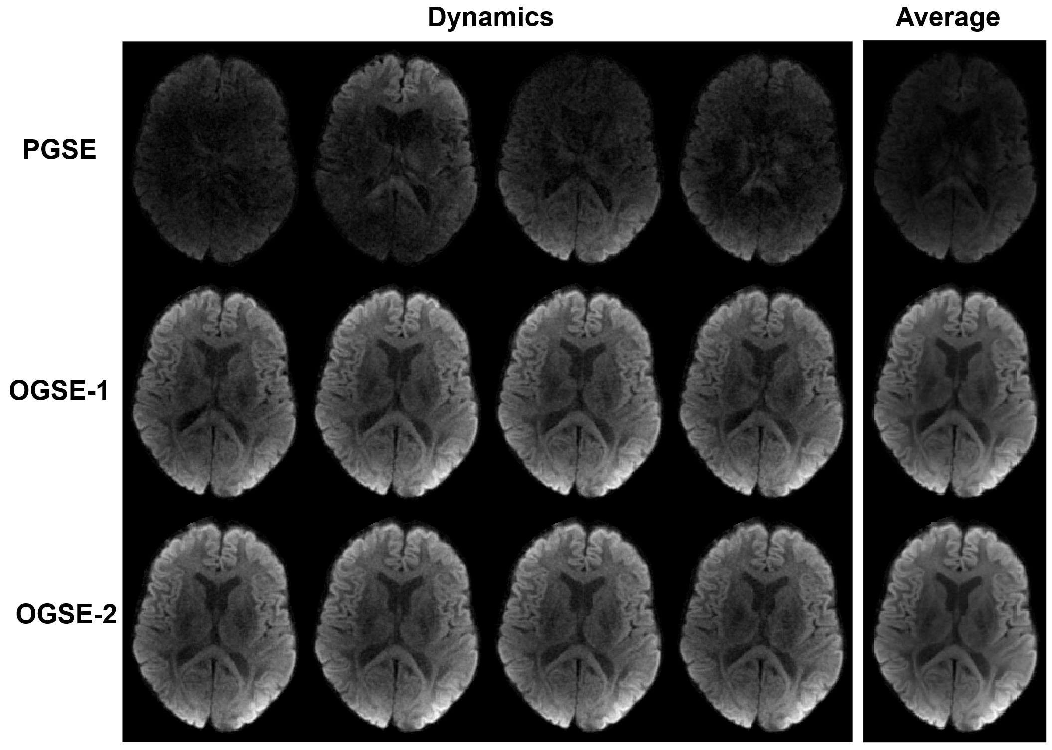
Figure 4. Time series of DW three-shot images across
dynamics for all gradient shapes (left), and the complex average across dynamics
(right). All images capture the same slice of one subject. The
diffusion-sensitizing gradient was aligned with the z-direction. Significant
artifacts confound each dynamic and the subsequent average for PGSE
acquisitions; no such issue occurs for either OGSE acquisition, for which fine
anatomical detail can be seen.

Figure 2. Phase differences with respect to the
first dynamic (different columns) across subsequent single-shot dynamics for
each diffusion sensitization scheme (different rows). Phase is observed to have
more pronounced fluctuations (i.e., of greater magnitude) among PGSE dynamics
than among dynamics of either OGSE acquisition. Between both forms of OGSE, phase
variations are similar.