Sofia Guterres1, Ana Rodrigues Fouto1, Nuno André Silva2, Pedro Vilela3, and Patrícia Figueiredo1
1Institute for Systems and Robotics - Lisboa and Department of Bioengineering, Instituto Superior Técnico, Universidade de Lisboa, Lisboa, Portugal, 2Hospital da Luz Learning Health, Lisboa, Portugal, 3Hospital da Luz, Lisboa, Portugal
1Institute for Systems and Robotics - Lisboa and Department of Bioengineering, Instituto Superior Técnico, Universidade de Lisboa, Lisboa, Portugal, 2Hospital da Luz Learning Health, Lisboa, Portugal, 3Hospital da Luz, Lisboa, Portugal
Simulations show that pCASL labelling efficiency depends on magnetic field inhomogeneity. In vivo data show no evidence of improved labelling efficiency or perfusion signal by using RF coil shimming of the labelling region.
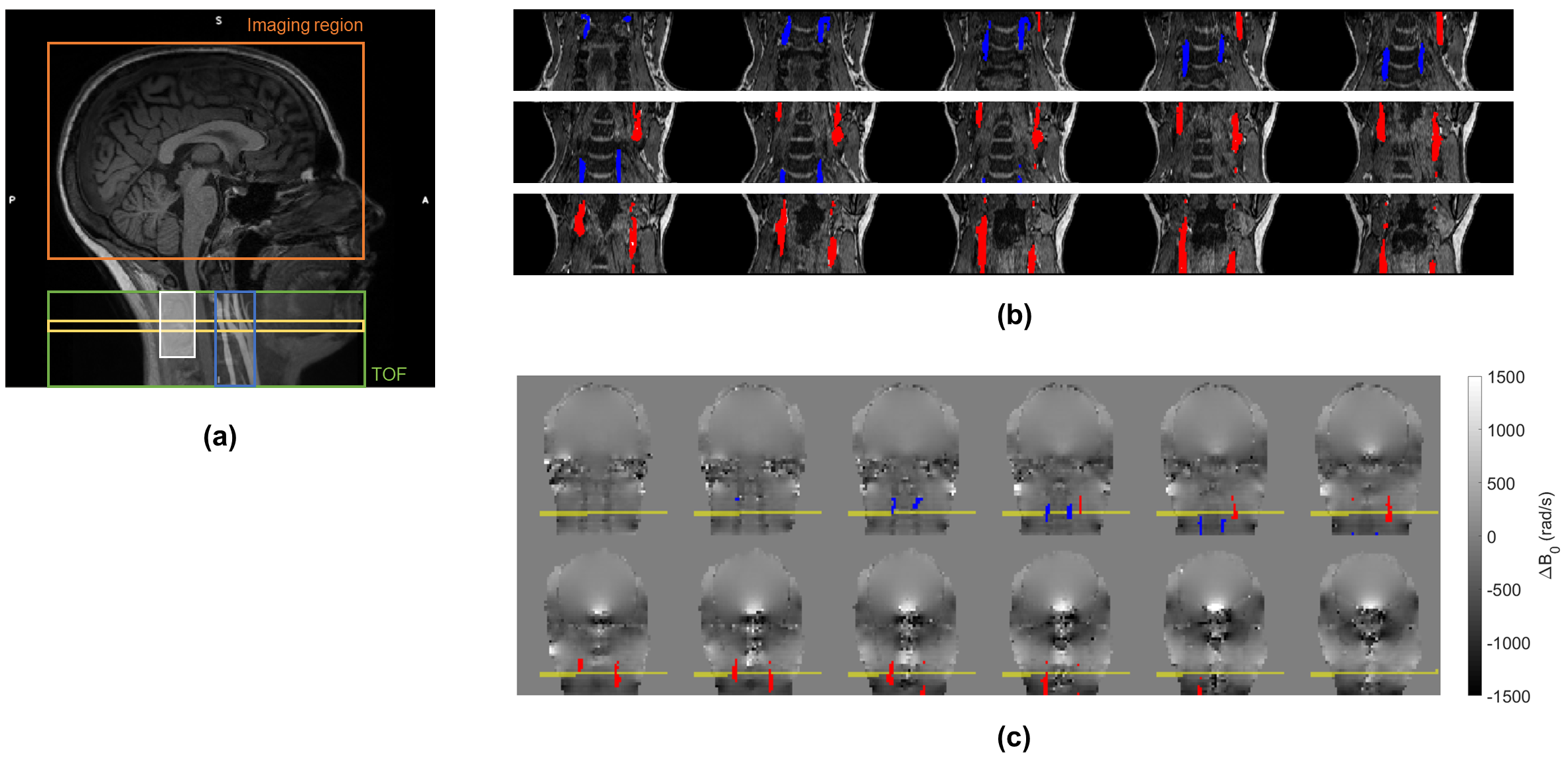
Fig. 1 - (a) Sagittal T1-weighted image indicating the pCASL imaging region (orange), TOF coverage (green), labelling plane (yellow), position of coronal slices for ROI definition as depicted in (b) (blue), and control region for field homogeneity (white); (b) TOF coronal slices showing the ICA (red) and VA (blue) ROIs; and (c) corresponding B0 fieldmap space (where the labelling plane is depicted in yellow), from which the ∆B0 values are extracted for the artery ROIs.
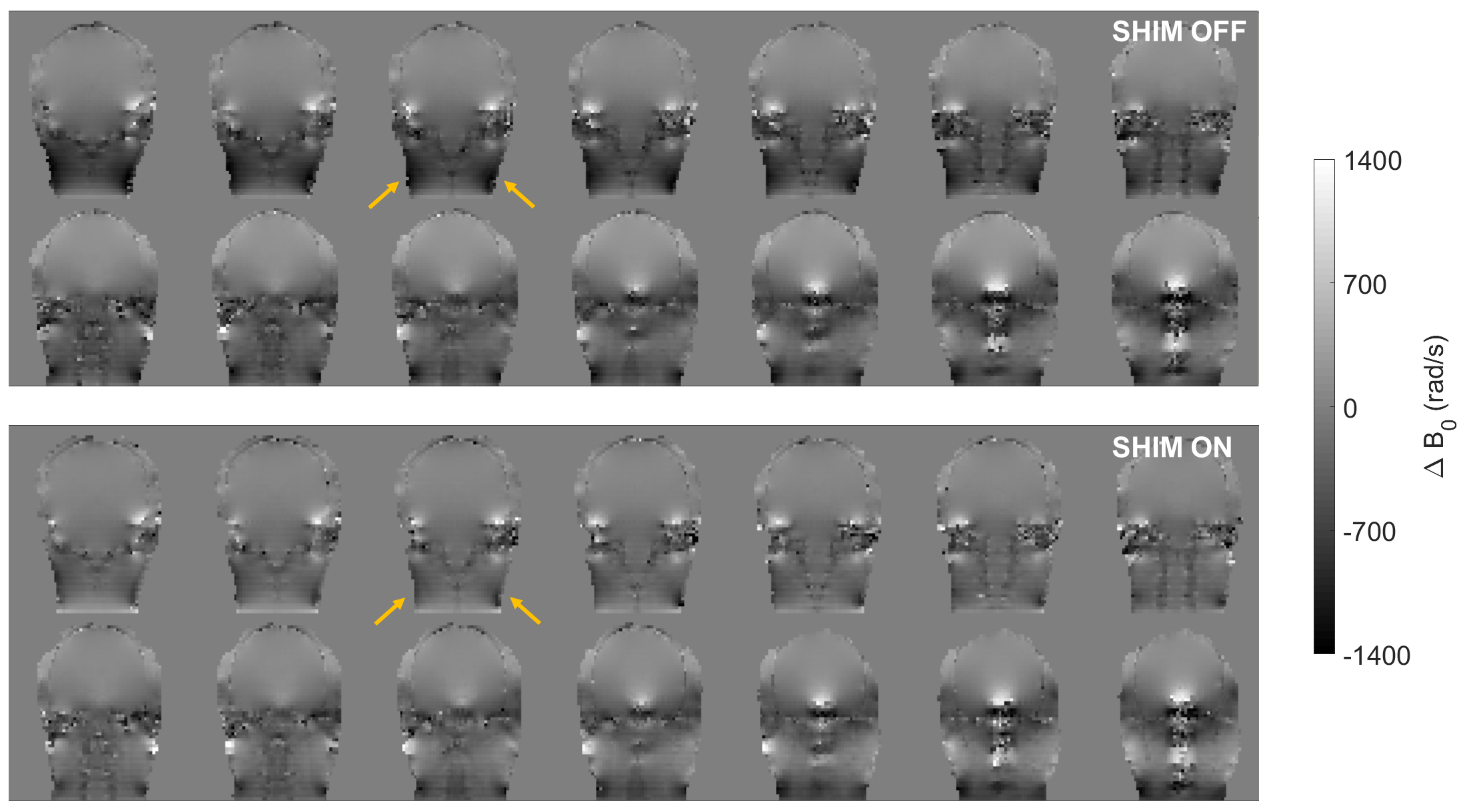
Fig. 3 - B0 fieldmap for an illustrative subject, with RF coil shiming “off” (top) and “on” (bottom). The reduction in field inhomogeneity between the shim “off” and “on” conditions is evident in the posterior neck region, as indicated by the arrows.