Matthäus Poniatowski1, Ilan Elias2, Mirsad Mahmutovic1, Gurinder Multani1, Sam-Luca J.D. Hansen1, Markus W. May1, Alexander M. König3, Jens H. Figiel3, Andreas H. Mahnken3, and Boris Keil1
1Institute of Medical Physics and Radiation Protection, TH Mittelhessen University of Applied Sciences, Gießen, Germany, 2Motionrad GmbH, Berlin, Germany, 3Department of Diagnostic and Interventional Radiology, Philipps-University Marburg, Marburg, Germany
1Institute of Medical Physics and Radiation Protection, TH Mittelhessen University of Applied Sciences, Gießen, Germany, 2Motionrad GmbH, Berlin, Germany, 3Department of Diagnostic and Interventional Radiology, Philipps-University Marburg, Marburg, Germany
To enable dynamic MRI for joints, an
in-bore motion-assisted device and a wearable coil array was designed,
constructed, and validated. The combination of robotic assisted joint motion, a
tight-fitting coil array, and accelerated imaging enabled dynamic MRI of the
angle.
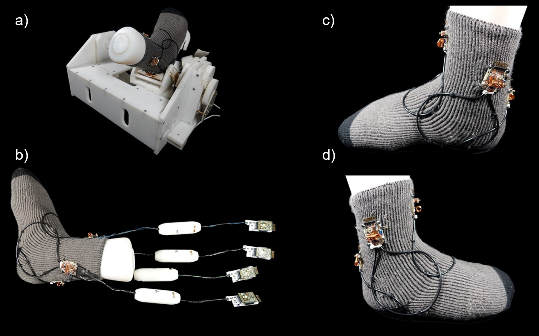
Figure 1: (a) 4-Channel High Impedance Coil placed on the
Robotic Motion Device. (b) Complete view of the 4ch HIC including transmission
lines, cable traps and preamplifiers. (c) Medial and (d) lateral close-up view
without transmission lines, cable traps and preamplifiers.

Figure
3: Comparison of SNR maps of
the HIC (top) and the LIC (bottom) using a foot-shaped agar phantom.