Angelika Mennecke1, Katrin Khakzar1, Kai Herz2, Moritz Fabian1, Alexander German1, Andrzej Liebert3, Ingmar Blümcke4, Burkhard Kasper5, Manuel Schmidt1, Arnd Dörfler1, Armin Nagel3, Frederik Laun3, and Moritz Zaiß1
1Department of Neuroradiology, University Hospital of Erlangen, FAU Erlangen-Nürnberg, Erlangen, Germany, 2High-field Magnetic Resonance Center, Max Planck Institute for Biological Cybernetics, Tübingen, Germany, 3Institute of Radiology, University Hospital of Erlangen, FAU Erlangen-Nürnberg, Erlangen, Germany, 4Institute of Neuropathology, University Hospital of Erlangen, FAU Erlangen-Nürnberg, Erlangen, Germany, 5Department of Neurology, Epilepsy Centre, University Hospital of Erlangen, FAU Erlangen-Nürnberg, Erlangen, Germany
1Department of Neuroradiology, University Hospital of Erlangen, FAU Erlangen-Nürnberg, Erlangen, Germany, 2High-field Magnetic Resonance Center, Max Planck Institute for Biological Cybernetics, Tübingen, Germany, 3Institute of Radiology, University Hospital of Erlangen, FAU Erlangen-Nürnberg, Erlangen, Germany, 4Institute of Neuropathology, University Hospital of Erlangen, FAU Erlangen-Nürnberg, Erlangen, Germany, 5Department of Neurology, Epilepsy Centre, University Hospital of Erlangen, FAU Erlangen-Nürnberg, Erlangen, Germany
With the help of the presented post-processing procedure, it is possible to
obtain an increased reproducibility. For the amide contrast, the CoV decreased from 4% to less than
1.2 %.

a) APT maps and b) NOE maps of a repeated measurement (group 1) before and after improving the post-processing steps (colorbar adjusted for visual comparison) c) decreasing CoV as a function of the stepwise improvement of post-processing methods: 1) moco with interpolation, 2) additionally, 2 point M0 normalization, 3) moco to the offset at 3.5 ppm, 4) B0 correction by using a Lorentzian fit 5) B0 correction with spline interpolation parameter 0.999 6) 2 point MZ normalization 7) LFM 4
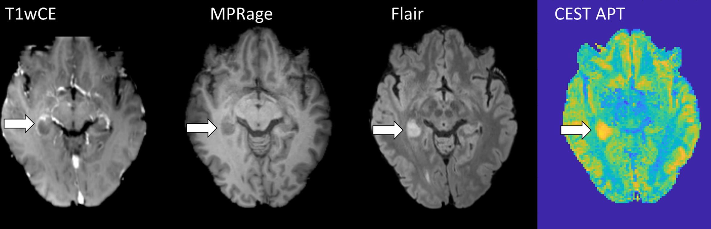
Conventional
images (contrast-enhanced T1 weighted imaging, MPRage, and Flair) and CEST APT
image of an epilepsy-associated tumor initially diagnosed as low grade,
histologically proven to be glioblastoma, IDH wild-type. Improved CEST postprocessing pipeline
was used.
