Huimin Zhang1, Baiyan Jiang1, Jian Hou1, Queenie Chan2, and Weitian Chen1
1Imaging and Interventional Radiology, The Chinese University of Hong Kong, Sha Tin, Hong Kong, 2Philips Healthcare, Hong Kong, Hong Kong
1Imaging and Interventional Radiology, The Chinese University of Hong Kong, Sha Tin, Hong Kong, 2Philips Healthcare, Hong Kong, Hong Kong
We utilized AC-iTIP and combined it with a proposed correction method to achieve robust quantification of T1rho.

Figure 3. a) A raw image acquired using AC-iTIP at TSL 0
ms. b) The fitting results from the selected ROI in a). c) The ground-truth T1rho
map acquired. d) and e) show the T1rho map before and after correction,
respectively. f) compares the T1rho values in c)-e). Blue, red and yellows bars
represent c), d) and e), respectively.
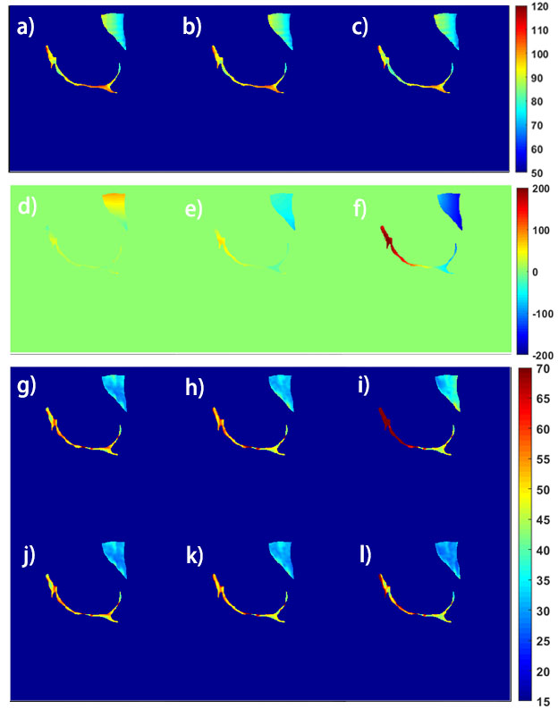
Figure 4. a)-c) and d)-e) show B1 and B0 field maps from three different scans. g)-i) and j)-l) show the T1rho maps in vivo before and after correction. The cartilage and muscle are manually drawn for analysis.
