Zhao Wei1,2,3, Alecio F. Lombardi1, Zubiad Ibrahim1, Mohammadamin Cheraghi1, Koihi Masuda4, Jiang Du1, Eric Y. Chang1,5, Graeme M. Bydder1, Wenhui Yang2,3, and Ya-Jun Ma1
1Department of Radiology, UC San Diego, San Diego, CA, United States, 2Institute of Electrical Engineering, Chinese Academy of Sciences, Beijing, China, 3University of Chinese Academy of Sciences, Beijing, China, 4Department of Orthopedic Surgery, UC San Diego, San Diego, CA, United States, 5Radiology Service, Veterans Affairs, San Diego Healthcare System, San Diego, CA, United States
1Department of Radiology, UC San Diego, San Diego, CA, United States, 2Institute of Electrical Engineering, Chinese Academy of Sciences, Beijing, China, 3University of Chinese Academy of Sciences, Beijing, China, 4Department of Orthopedic Surgery, UC San Diego, San Diego, CA, United States, 5Radiology Service, Veterans Affairs, San Diego Healthcare System, San Diego, CA, United States
The
UTE-Adiab-T1ρ technique can quantify
the T1ρ
of whole lumbar IVDs, including the nucleus pulposus,
annulus fibrosis and cartilaginous endplate. This may be valuable for comprehensive
assessment of IVD degeneration.
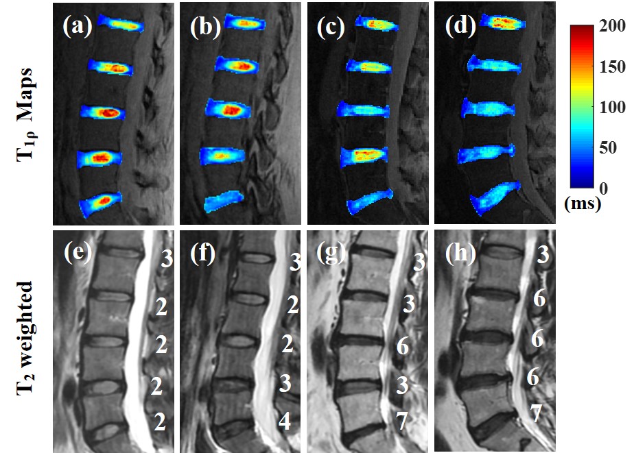
Figure 2. Representative T1ρ maps (first row)
and corresponding T2w-FSE images (second row) of four subjects (as
shown in four columns respectively). These T1ρ maps demonstrate that
UTE-Adiab-T1ρ sequence allows measurement of T1ρs of
the whole IVD, including the CEP. In comparison, the
CEP regions were low signal on the T2w-FSE images. The modified Pfirrmann
grades of discs are included on the T2w-FSE images.
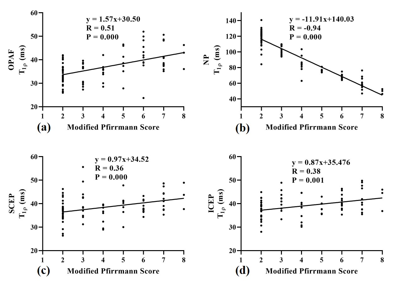
Figure 3.
Spearman’s correlation coefficient results between modified Pfirrmann grades
and T1ρ values of OPAF (a), SCEP (b), ICEP (c),
and NP (d). A strong negative correlation is observed in the NP, and moderate
positive correlations are observed in the OPAF, SCEP, and ICEP.