Ya Guo1, Haihua Bao1, and Shaoyu Wang2
1Affiliated Hospital of Qinghai University, Xining, China, 2Siemens Healthineers, Shanghai, China
1Affiliated Hospital of Qinghai University, Xining, China, 2Siemens Healthineers, Shanghai, China
The differences in brain function and functional
connection provide theoretical support for the timely treatment of vestibular
migraine in clinical practice.
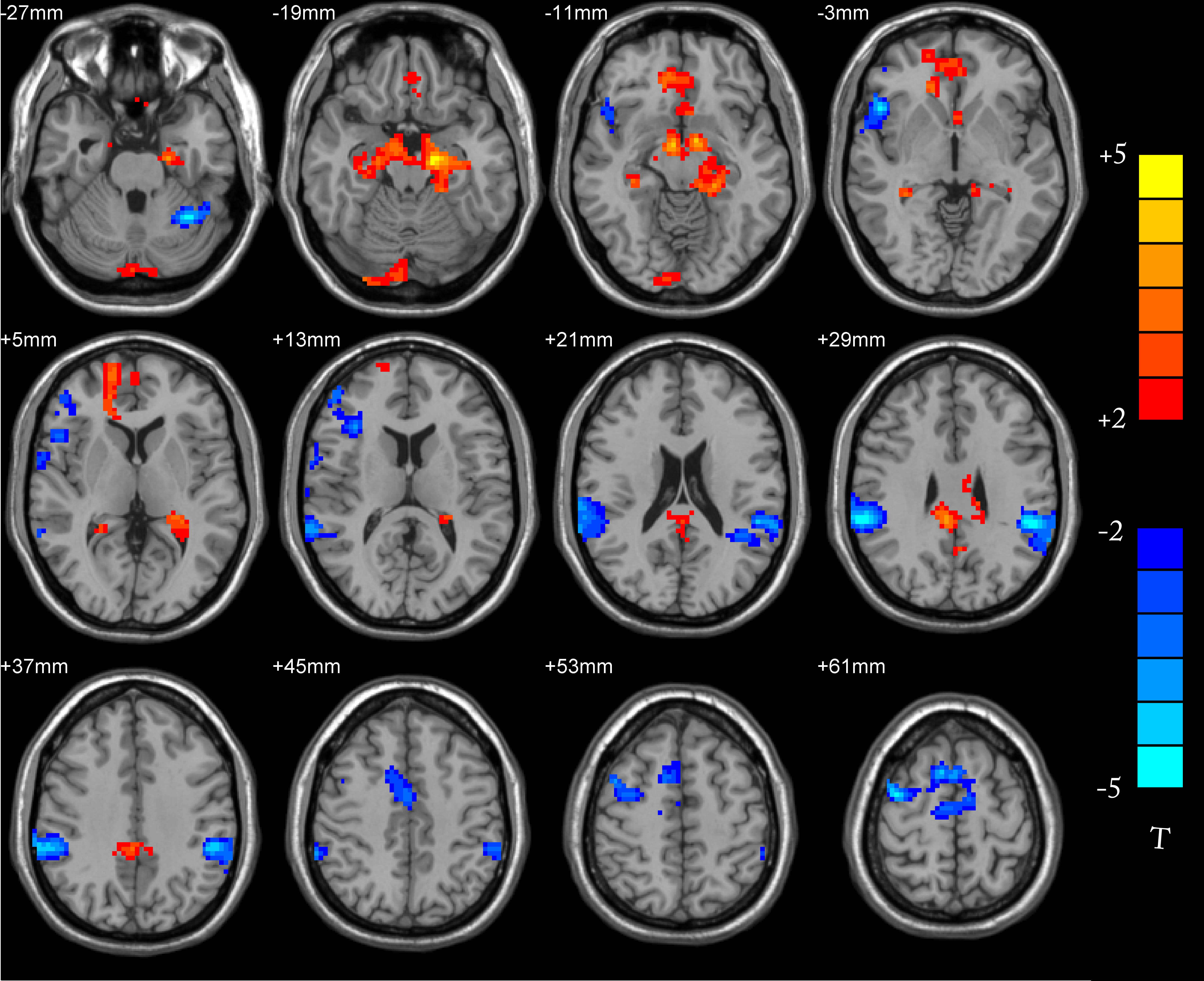
Compared with the normal group, the brain areas with enhanced
functional connection between DMN and PCC in the case group were red-yellow,
and the brain areas with weakened connection were blue.
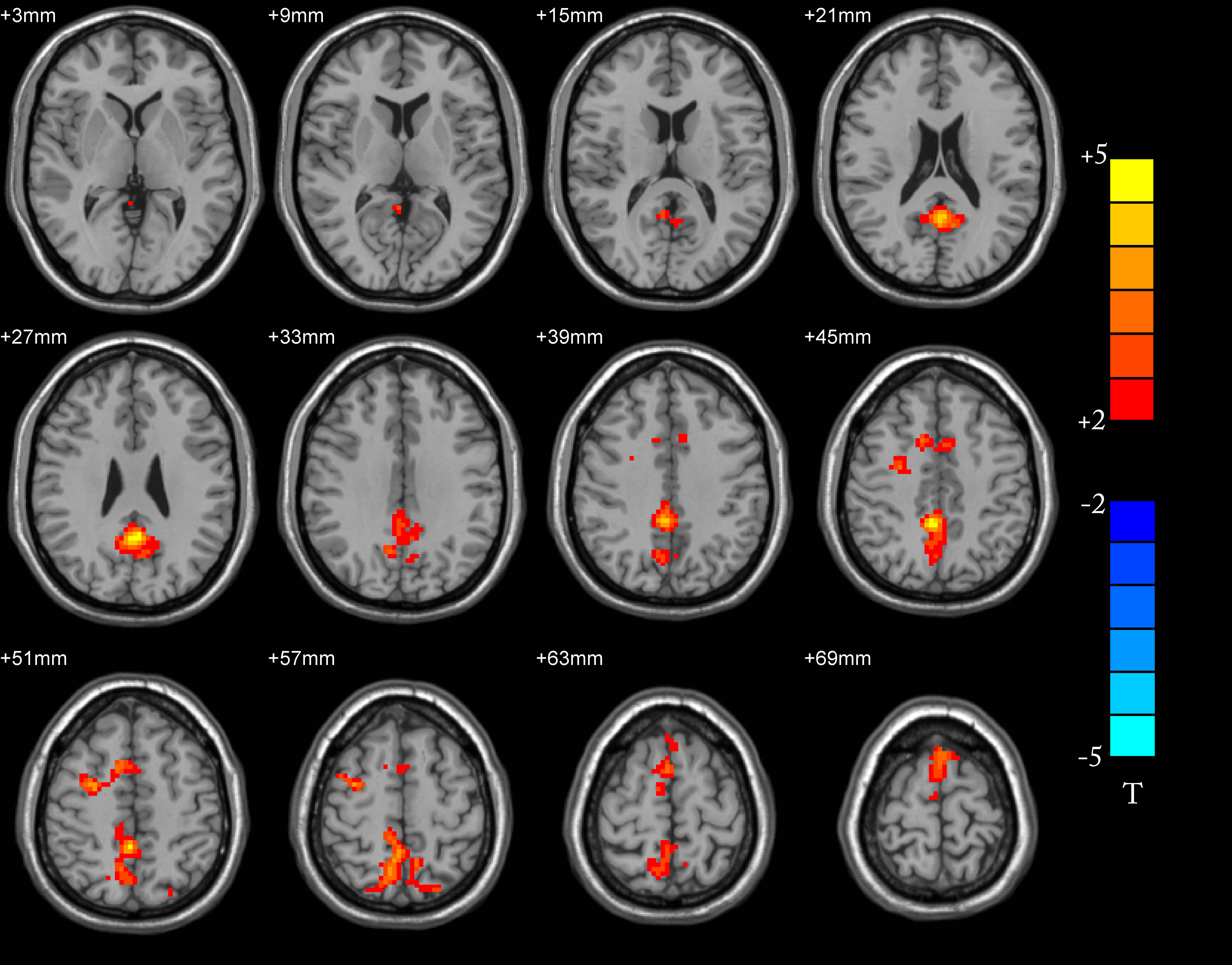
The ALFF values about patient group were increased in bilateral
anterior cuneiform lobes, bilateral medial and lateral cingulate gyrus,
bilateral posterior cingulate gyrus, bilateral supplementary motor areas,
bilateral dorsolateral superior frontal gyrus, left cuneiform lobe, right In
the lateral middle frontal lobe, the right central anterior gyrus, the left
paracentral lobule and the left medial superior frontal gyrus, there were no
brainareas with reduced ALFF values in the whole brain.