Jo Lee1,2, Sen Jia1,2, Liu Liu3, Xiaoliang Zhang4, and Ye Li1,2
1Paul C. Lauterbur Research Center for Biomedical Imaging, Shenzhen Institutes of Advanced Technology, Chinese Academy of Sciences, Shenzhen, China, 2Shenzhen Key Laboratory for MRI, Shenzhen, China, 3United Imaging Healthcare, Shanghai, China, 4Department of Biomedical Engineering, State University of New York, Buffalo, NY, United States
1Paul C. Lauterbur Research Center for Biomedical Imaging, Shenzhen Institutes of Advanced Technology, Chinese Academy of Sciences, Shenzhen, China, 2Shenzhen Key Laboratory for MRI, Shenzhen, China, 3United Imaging Healthcare, Shanghai, China, 4Department of Biomedical Engineering, State University of New York, Buffalo, NY, United States
We designed a quadrature-birdcage/ 47Rx head coil array
for accelerated images on 3 T MRI, and has compared to a commercial 32-channel head
coil for quantified analysis. The results show that the 47-channel head coil has
better acceleration ability.
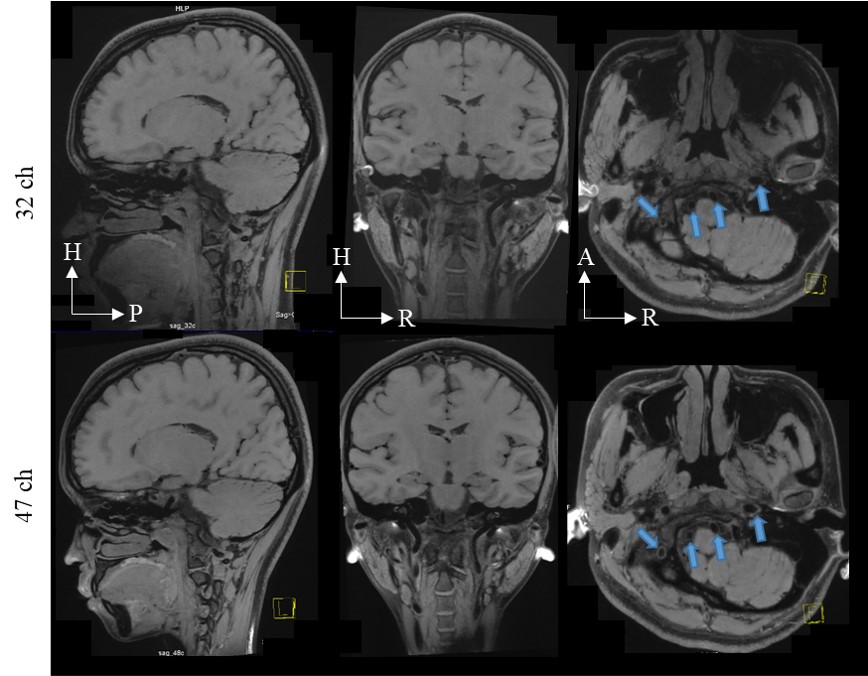
Fig.3. 0.6 mm isotropic modulated
flip angle technique in refocused imaging with extended echo train (MATRIX)
images were acquired using the commercial head coil (32ch; top row) and the quadrature
birdcage/47Rx coil array (47ch; bottom row) at R = 7-fold acceleration. The
acceleration direction was on phase encoding and slice phase encoding. H =
head; P = posterior; R = right; A = anterior.
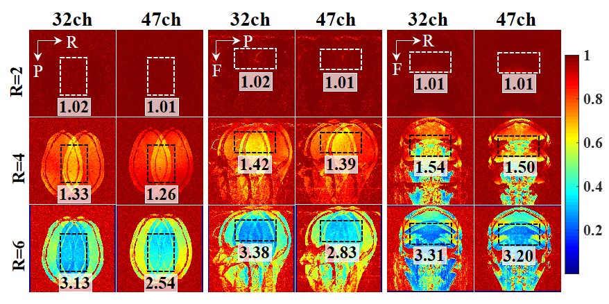
Fig.2. Inverse g-factor maps. To
display coil acceleration ability of each direction, each orientation was
accelerated on different directions. Transverse orientation: Left-to-Right
acceleration direction. Sagittal orientation: Anterior-to-Posterior
acceleration direction. Coronal orientation: Head-to-Feet acceleration
direction. Regions of interest (ROIs) is marked as black dashed line. The mean
g-factor value of each ROI is written beside the related image. L: left, R:
right, A: anterior, P: posterior, H: head, F: feet.