Ilias Giannakopoulos1, Georgy Dmitrievich Guryev2, Jose Enrique Cruz Serralles2, Ioannis Georgakis1, Luca Daniel2, Jacob White2, and Riccardo Lattanzi1,3,4
1Center for Advanced Imaging Innovation and Research (CAI2R), Department of Radiology, New York University Grossman School of Medicine, New York, NY, United States, 2Department of Electrical & Computer Engineering, Massachusetts Institute of Technology, Cambridge, MA, United States, 3The Bernard and Irene Schwartz Center for Biomedical Imaging (CBI), Department of Radiology, New York University Grossman School of Medicine, New York, NY, United States, 4Vilcek Institute of Graduate Biomedical Sciences, New York University Grossman School of Medicine, New York, NY, United States
1Center for Advanced Imaging Innovation and Research (CAI2R), Department of Radiology, New York University Grossman School of Medicine, New York, NY, United States, 2Department of Electrical & Computer Engineering, Massachusetts Institute of Technology, Cambridge, MA, United States, 3The Bernard and Irene Schwartz Center for Biomedical Imaging (CBI), Department of Radiology, New York University Grossman School of Medicine, New York, NY, United States, 4Vilcek Institute of Graduate Biomedical Sciences, New York University Grossman School of Medicine, New York, NY, United States
The volume-surface integral equation coupling matrix,
that models the interactions between radiofrequency coils and tissue, is reshaped to a set of incomplete tensors and
compressed with the canonical model. Results show 35 times compression for an error
less than 2%, at a 7 T MRI simulation.
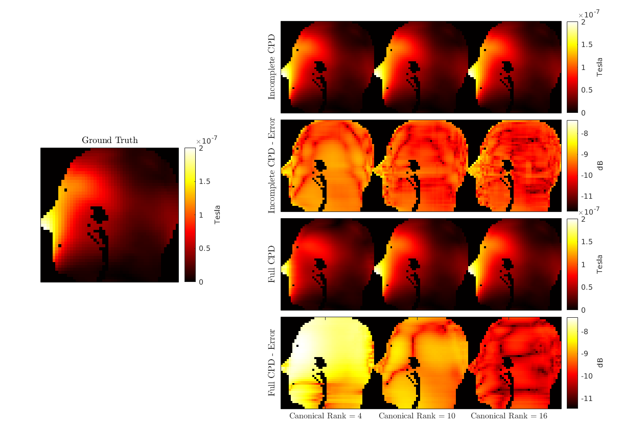
Figure
4: Qualitative comparison
between the simulated
B1+ maps,
for
one representative coil
element of the array and the
middle sagittal slice. The
results for the incomplete and full matrix are displayed for
three canonical ranks (4,10,16), along
with the relative error with respect to the ground truth (left)
in logarithmic scale. Voxels
outside the scatterer are masked for enhanced visualization.

Figure
3: Overall compression factor
(left axis) and relative error (right axis), for the full and
incomplete matrices and various canonical ranks.
