Peiyao Chen1, Chao Jin1, Xianjun Li1, Miaomiao Wang1, Congcong Liu1, Xiaoyu Wang1, Fan Wu1, Yuli Zhang1, Cong Tian1, Mengxuan Li1, Xiaocheng Wei2, and Jian Yang1
1First Affiliated Hospital of Xi 'an Jiaotong University, Xi'an, Shaanxi, China, 2MR Research China, GE Healthcare, Beijing, China
1First Affiliated Hospital of Xi 'an Jiaotong University, Xi'an, Shaanxi, China, 2MR Research China, GE Healthcare, Beijing, China
Preterm
children showed lower cerebral blood flow than term in the frontal lobe, even
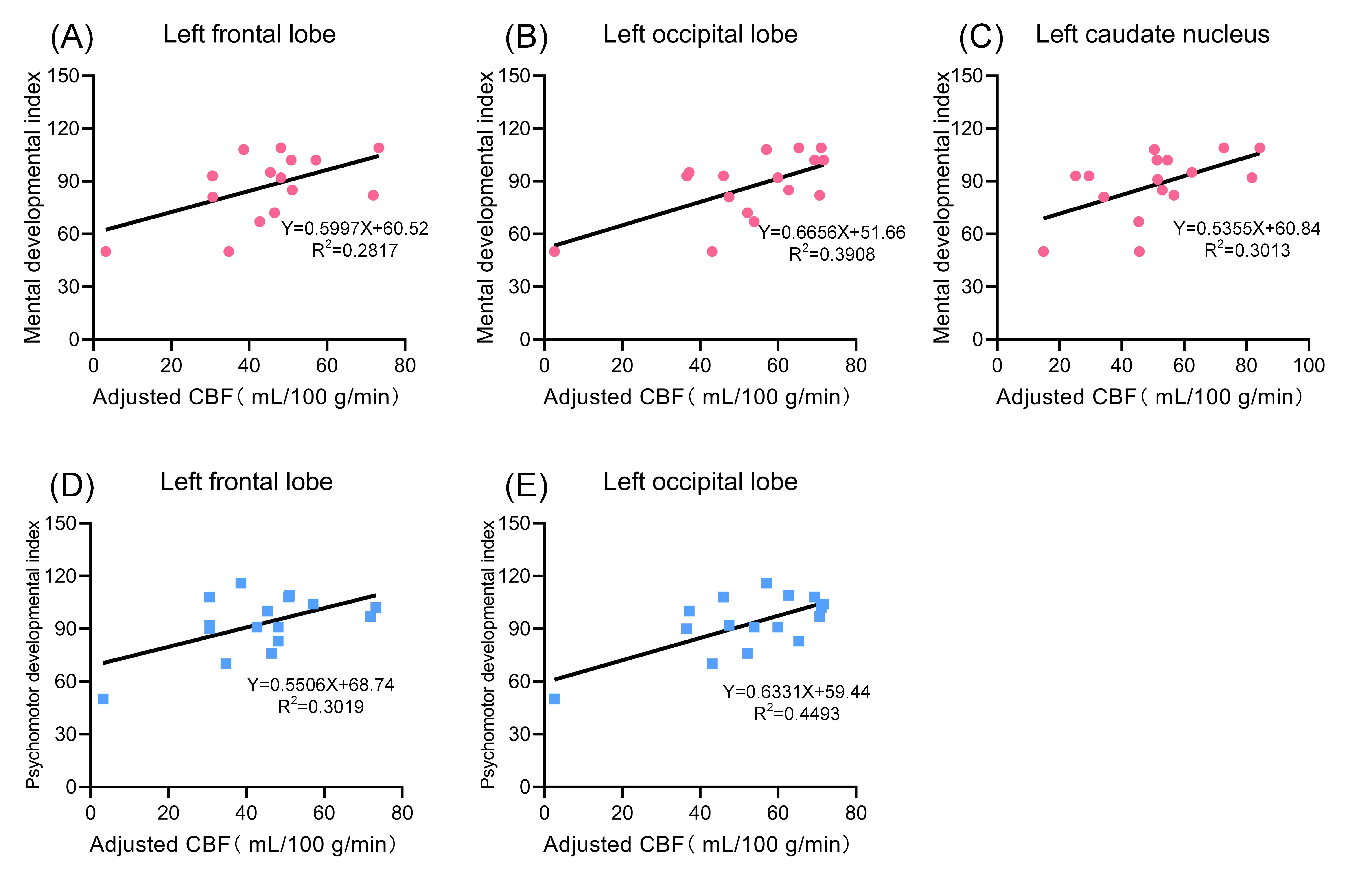
at late childhood. After controlling for age, CBF showed positive correlations with
mental and psychomotor development indexes in left frontal and occipital lobes
in preterm children.
Figure
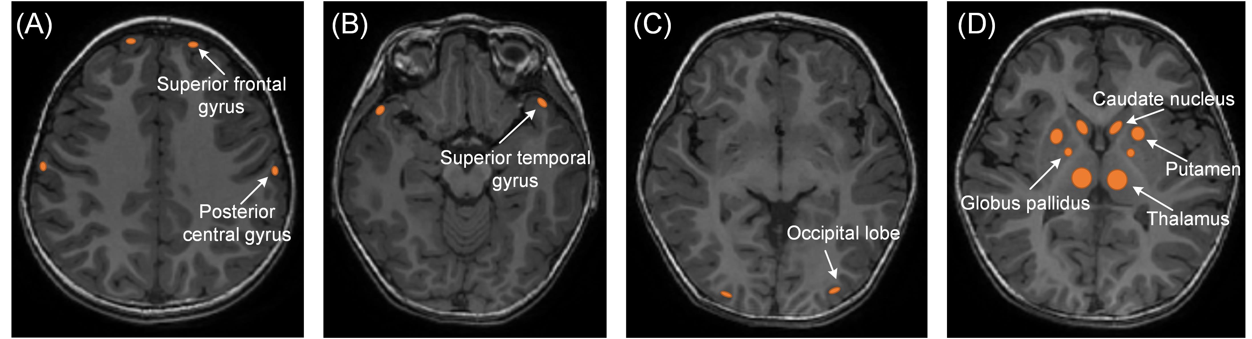
1. Manual regions of interest (ROIs) were placed on the CBF map by using the
aligned anatomical image as guidance. ROI was about 20-100mm2. (A)
bilateral superior frontal gyrus and posterior central gyrus.(B)bilateral
superior temporal gyrus.(C)bilateral occipital lobe.(D)basal ganglia (bilateral
thalamus, globus pallidus, putamen, caudate nucleus)
Figure
4. Regions of positive correlation between MDI、PDI
and CBF in preterm infants aged 60 days-4 years of age. MDI (pink): left
frontal、occipital lobe and
caudate nuclear. PDI (blue): left frontal and occipital lobe.