Douglas C Dean1,2,3, Nagesh Adluru3, Jace B King4, Molly B Prigge4, Carolyn King4, Erin D Bigler5,6,7,8, June Taylor4, Nick Lange9, Brandon A Zielinski4,5,10, Janet E Lainhart3,11, and Andrew L Alexander2,3,11
1Pediatrics, University of Wisconsin–Madison, Madison, WI, United States, 2Medical Physics, University of Wisconsin–Madison, Madison, WI, United States, 3Waisman Center, University of Wisconsin–Madison, Madison, WI, United States, 4Radiology, University of Utah, Salt Lake City, UT, United States, 5Neurology, University of Utah, Salt Lake City, UT, United States, 6Psychiatry, University of Utah, Salt Lake City, UT, United States, 7Psychology and Neuroscience Center, Brigham Young University, Provo, UT, United States, 8Neurology, University of California–Davis, Davis, CA, United States, 9Psychiatry, Harvard School of Medicine, Boston, MA, United States, 10Pediatrics, University of Utah, Salt Lake City, UT, United States, 11Psychiatry, University of Wisconsin–Madison, Madison, WI, United States
1Pediatrics, University of Wisconsin–Madison, Madison, WI, United States, 2Medical Physics, University of Wisconsin–Madison, Madison, WI, United States, 3Waisman Center, University of Wisconsin–Madison, Madison, WI, United States, 4Radiology, University of Utah, Salt Lake City, UT, United States, 5Neurology, University of Utah, Salt Lake City, UT, United States, 6Psychiatry, University of Utah, Salt Lake City, UT, United States, 7Psychology and Neuroscience Center, Brigham Young University, Provo, UT, United States, 8Neurology, University of California–Davis, Davis, CA, United States, 9Psychiatry, Harvard School of Medicine, Boston, MA, United States, 10Pediatrics, University of Utah, Salt Lake City, UT, United States, 11Psychiatry, University of Wisconsin–Madison, Madison, WI, United States
Main findings highlight significant microstructural alterations of the corpus callosum in ASD using advanced diffusion imaging and quantitative relaxometry.
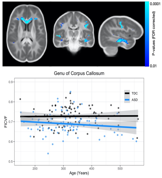
Figure 1: Significantly lower NODDI NDI found in ASD compared to TDC (p<0.05, corrected). Lower NDI were observed in areas of the genu of the corpus callosum, superior longitudinal fasciculus, and uncinate fasciculus.

Figure 2: Significant age by group interactions of qT1 between ASD and TDC subjects overlaid on the mean qT1 map. Significant interactions were localized in the in body and splenium of the corpus callosum.